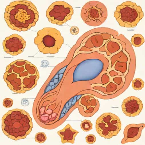
Adrenal cortical adenomas are a common type of benign tumor found in the adrenal cortex, often discovered incidentally during imaging for unrelated conditions. Understanding their pathology is crucial for accurate diagnosis, treatment planning, and differentiation from malignant adrenal tumors. These adenomas can be hormonally active, leading to clinical syndromes, or non-functional and asymptomatic. Examining adrenal cortical adenoma pathology outlines allows medical professionals, students, and researchers to grasp the cellular, morphological, and clinical features of these tumors, which is essential for effective patient management and research advancement.
Definition and General Features
An adrenal cortical adenoma is a benign neoplasm originating from the cells of the adrenal cortex. These tumors are usually small, well-circumscribed, and often unilateral. The adrenal cortex consists of three zones the zona glomerulosa, zona fasciculata, and zona reticularis, each responsible for producing different steroid hormones. Adenomas may arise from any of these zones, influencing the type of hormones secreted and the clinical manifestations.
Key Pathological Features
- Well-circumscribed, encapsulated nodules often less than 5 cm in diameter.
- Composed of uniform, polygonal cells with abundant cytoplasm.
- Low mitotic activity and absence of significant atypia or necrosis.
- May contain lipid-rich cells, giving a yellowish appearance on gross examination.
- Usually unilateral but can rarely be bilateral.
Understanding these basic features is fundamental for distinguishing adenomas from adrenal cortical carcinomas and other adrenal lesions.
Histological Characteristics
Histologically, adrenal cortical adenomas exhibit distinct cellular patterns depending on the originating cortical zone. Cells are arranged in nests, cords, or trabeculae, separated by delicate vascular stroma. The cytoplasm can be clear or eosinophilic due to the presence of lipids, which are essential in steroid hormone synthesis. Nuclei are typically round with fine chromatin and inconspicuous nucleoli. Mitotic figures are rare, and atypical mitoses are generally absent, distinguishing adenomas from malignant counterparts.
Zone-Specific Features
- Zona glomerulosa adenomasMay produce aldosterone, leading to primary hyperaldosteronism.
- Zona fasciculata adenomasOften secrete cortisol, causing Cushing’s syndrome.
- Zona reticularis adenomasCan produce androgens, sometimes resulting in virilization.
Recognizing the zone of origin helps pathologists predict the functional status of the adenoma and potential clinical manifestations.
Functional vs. Non-Functional Adenomas
Adrenal cortical adenomas can be classified based on hormonal activity. Functional adenomas secrete excess hormones, leading to distinct clinical syndromes, whereas non-functional adenomas do not produce significant hormones and are often discovered incidentally on imaging studies. Functional adenomas require careful biochemical evaluation to identify hormone excess and guide treatment.
Clinical Syndromes Associated with Functional Adenomas
- Primary hyperaldosteronismCaused by aldosterone-secreting adenomas, leading to hypertension and hypokalemia.
- Cushing’s syndromeResulting from cortisol-secreting adenomas, characterized by weight gain, central obesity, and metabolic disturbances.
- Virilization or feminizationDue to androgen or estrogen-secreting adenomas, causing changes in secondary sexual characteristics.
Identifying the functional status of an adrenal cortical adenoma is essential for deciding between surgical and medical management.
Gross Pathology
On gross examination, adrenal cortical adenomas are usually small, ranging from 1 to 5 cm, although larger tumors can occur. They are well-demarcated, encapsulated, and cut surfaces often appear yellow due to lipid content. Hemorrhage or necrosis is uncommon in benign adenomas, and the adrenal gland surrounding the tumor generally appears normal. These features are critical for pathologists to distinguish adenomas from more aggressive adrenal lesions.
Important Gross Features
- Size typically less than 5 cm in diameter.
- Well-circumscribed and encapsulated.
- Yellow cut surface due to lipid-rich cytoplasm.
- Minimal or no hemorrhage and necrosis.
- Unilateral predominance, although bilateral adenomas can occur.
Diagnostic Workup
Diagnosis of adrenal cortical adenomas involves a combination of imaging, biochemical testing, and histopathological evaluation. Imaging techniques such as CT and MRI help identify the size, location, and characteristics of the lesion. Lipid-rich adenomas typically show low attenuation on CT, whereas MRI can provide additional information about tissue composition. Biochemical testing assesses hormone levels to determine whether the adenoma is functional. Ultimately, histopathological examination confirms the diagnosis and rules out malignancy.
Diagnostic Steps
- Imaging CT or MRI to evaluate size, morphology, and lipid content.
- Biochemical tests Plasma and urine hormone levels to assess functionality.
- Histology Microscopic examination to evaluate cell type, arrangement, and atypia.
- Immunohistochemistry Used in certain cases to differentiate adenomas from carcinomas.
A thorough diagnostic workup ensures accurate identification and appropriate management of adrenal cortical adenomas.
Treatment and Prognosis
Treatment for adrenal cortical adenomas depends on size, hormonal activity, and clinical symptoms. Functional adenomas causing significant hormone excess typically require surgical removal, often via laparoscopic adrenalectomy. Non-functional adenomas may be monitored with periodic imaging if they are small and asymptomatic. Prognosis for benign adenomas is excellent, with minimal risk of recurrence after complete resection.
Management Guidelines
- Functional adenomas Surgical removal to alleviate hormone excess and associated symptoms.
- Non-functional adenomas<4 cm Often monitored with periodic imaging and follow-up.
- Non-functional adenomas >4 cm Consider surgical excision due to increased risk of malignancy.
- Postoperative monitoring Assess hormone levels and imaging to ensure no recurrence.
Appropriate management and follow-up are essential to maintain patient health and prevent complications associated with functional adenomas.
Adrenal cortical adenomas are benign tumors that arise from the adrenal cortex, with distinct pathological, histological, and clinical features. Understanding their pathology through outlines of gross and microscopic characteristics helps distinguish them from malignant tumors and guides appropriate clinical management. Functional adenomas can lead to specific hormonal syndromes, while non-functional adenomas are often incidental findings. Accurate diagnosis, using imaging, biochemical tests, and histology, is essential for optimal treatment planning. Overall, adrenal cortical adenomas generally have an excellent prognosis, and knowledge of their pathology is crucial for medical professionals and students seeking to understand adrenal diseases and endocrine tumors.