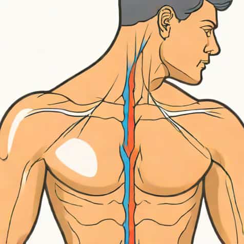
The trapezius is one of the largest and most superficial muscles of the back and neck, playing a crucial role in the movement and stabilization of the shoulder girdle and upper spine. Understanding its innervation is essential for medical students, physiotherapists, and healthcare professionals, as it directly impacts clinical assessments, surgical approaches, and rehabilitation strategies. The trapezius muscle’s unique innervation allows it to perform diverse functions, from shrugging the shoulders to extending the head, making it a critical component of human anatomy and motor function.
Overview of the Trapezius Muscle
The trapezius is a broad, flat, triangular-shaped muscle that spans the posterior thoracic and cervical regions. It is divided into three functional regions the upper, middle, and lower fibers, each contributing to specific movements of the scapula and neck. The upper fibers elevate the scapula and assist in neck extension, the middle fibers retract the scapula, and the lower fibers depress the scapula and support upward rotation. The wide range of functions of the trapezius makes its innervation particularly significant in clinical and anatomical contexts.
Anatomical Attachments
The trapezius originates from the occipital bone, the ligamentum nuchae, and the spinous processes of the seventh cervical and all thoracic vertebrae. It inserts onto the lateral third of the clavicle, the acromion, and the spine of the scapula. These attachments allow the muscle to exert force over the shoulder girdle and upper back, influencing posture and coordinated arm movements. Any compromise in its innervation can lead to significant functional deficits, including winged scapula or limited shoulder mobility.
Innervation of the Trapezius
The trapezius muscle is primarily innervated by the accessory nerve, also known as cranial nerve XI, which provides motor input to enable contraction of the muscle fibers. In addition to the accessory nerve, the trapezius receives sensory fibers from the cervical plexus, particularly the ventral rami of C3 and C4, which contribute proprioceptive feedback and assist in muscle tone regulation. This dual innervation-motor from cranial nerve XI and sensory from cervical spinal nerves-is essential for coordinated movements and postural control.
The Accessory Nerve (Cranial Nerve XI)
The accessory nerve is a motor nerve that arises from the medulla oblongata and upper cervical spinal cord. It descends through the jugular foramen and travels along the lateral aspect of the neck to reach the trapezius. The nerve enters the muscle at its posterior border and branches extensively to supply all three regions of the trapezius. Damage to the accessory nerve, whether due to trauma, surgery, or neurological conditions, can result in weakness or paralysis of the trapezius, leading to drooping shoulders, difficulty in shoulder elevation, and impaired arm abduction above the horizontal plane.
Cervical Plexus Contribution
While the accessory nerve provides motor innervation, the sensory input to the trapezius comes from the cervical plexus, mainly the C3 and C4 spinal nerves. These nerves provide proprioceptive information about muscle stretch, tension, and position, enabling the central nervous system to coordinate precise movements and maintain posture. This sensory feedback is crucial during complex activities such as lifting, reaching, or rotating the head and neck. Lesions affecting the cervical plexus can compromise proprioception and result in abnormal movement patterns or postural instability.
Functions of the Trapezius Related to Innervation
The coordinated innervation of the trapezius allows it to perform multiple essential functions, including scapular elevation, retraction, and rotation, as well as neck extension and lateral flexion. Proper functioning of the accessory nerve ensures that motor signals reach all regions of the trapezius, while sensory input from C3 and C4 enables feedback mechanisms that modulate strength and coordination. Together, these innervations allow the trapezius to support both dynamic movements and static postural adjustments.
Movements Facilitated by the Trapezius
- Scapular ElevationRaising the shoulder girdle, essential for shrugging.
- Scapular RetractionPulling the shoulder blades toward the spine.
- Scapular DepressionLowering the scapula, which stabilizes the shoulder during arm movements.
- Upward RotationFacilitates arm abduction above the head.
- Neck Extension and Lateral FlexionControlled by upper fibers during head movements.
Clinical Significance of Trapezius Innervation
Understanding the innervation of the trapezius is critical in clinical practice. Injury to the accessory nerve is a common complication during surgical procedures such as lymph node dissection in the posterior triangle of the neck. Patients with accessory nerve injury often present with shoulder droop, winged scapula, and difficulty raising the arm above the horizontal plane. Electromyography and nerve conduction studies can help evaluate the functional status of the trapezius and guide rehabilitation. In addition, physical therapy interventions often target strengthening the trapezius and compensating for any deficits caused by nerve injury.
Common Conditions Affecting Trapezius Innervation
- Accessory nerve injury during neck surgery or trauma.
- Cervical plexus lesions due to spinal injury or compression.
- Neuromuscular diseases such as amyotrophic lateral sclerosis or peripheral neuropathies.
- Postural disorders leading to trapezius strain or chronic tension.
Testing Trapezius Function
Evaluating trapezius function involves both clinical examination and neurological testing. Clinicians often ask patients to shrug their shoulders against resistance to assess upper fiber strength, and they observe scapular positioning for signs of paralysis or weakness. Sensory input from C3 and C4 can be tested indirectly by assessing proprioception and coordination of shoulder and neck movements. Accurate assessment of trapezius innervation is important for diagnosing nerve injuries and planning effective rehabilitation strategies.
Physical Examination Techniques
- Manual muscle testing for shoulder elevation and abduction.
- Observation of scapular asymmetry or winging.
- Palpation of the trapezius during contraction.
- Assessment of neck movements such as extension and lateral flexion.
Rehabilitation and Treatment Approaches
Rehabilitation of trapezius dysfunction depends on the underlying cause. Physical therapy often focuses on strengthening the remaining functional fibers, improving posture, and restoring mobility. Electrical stimulation may be used in cases of partial accessory nerve injury to enhance muscle activation. In severe cases, surgical nerve repair or tendon transfer may be necessary to restore function. Understanding the innervation pathways is essential for designing effective interventions and ensuring optimal recovery of muscle strength and coordination.
Exercise Recommendations
- Shoulder shrugs to strengthen upper fibers.
- Scapular retraction exercises for middle fibers.
- Prone arm lifts and scapular depression exercises for lower fibers.
- Postural training and proprioceptive exercises to support cervical plexus function.
The innervation of the trapezius is a critical component of its ability to control the shoulder and neck. The accessory nerve (cranial nerve XI) provides motor input, while the cervical plexus (C3 and C4) supplies sensory and proprioceptive feedback. This dual innervation allows the trapezius to perform complex movements, maintain posture, and contribute to both dynamic and static functions of the upper back and neck. Knowledge of its innervation is essential for clinical assessment, diagnosis of nerve injuries, surgical planning, and rehabilitation strategies. Understanding how the trapezius is innervated not only clarifies its functional capabilities but also highlights the importance of preserving nerve integrity for overall musculoskeletal health.