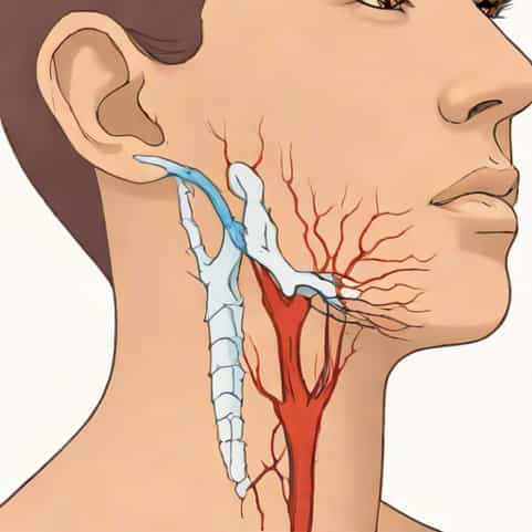
Jugular vein distention is a visible enlargement of the jugular veins in the neck, often indicating an underlying cardiovascular or systemic problem. Understanding the pathophysiology of jugular vein distention is crucial because it reflects abnormal pressure changes within the venous system, especially in the right side of the heart. When the jugular veins appear prominent or bulging, it is not merely a cosmetic observation but a clinical sign pointing to hemodynamic disturbances. By exploring the mechanisms, causes, and physiological processes involved, healthcare professionals and patients can better recognize the significance of this finding and seek appropriate evaluation and treatment.
Anatomy and Normal Function of the Jugular Veins
The jugular veins, which include the internal and external jugular veins, are major vessels responsible for draining blood from the head and neck back to the heart. Under normal conditions, these veins collapse when a person is upright because venous pressure is low and blood flows freely toward the right atrium. Observing the jugular venous pulse can give valuable insights into right atrial pressure and central venous pressure, making it an important clinical tool during physical examination.
In healthy individuals, the jugular veins are barely visible when sitting at a 45-degree angle. Any distention suggests an elevation in central venous pressure or an obstruction to venous return, which sets the stage for understanding the pathophysiology of jugular vein distention.
Pathophysiology of Jugular Vein Distention
The pathophysiology of jugular vein distention primarily involves increased pressure within the venous system. This elevation in pressure occurs when the right atrium or right ventricle fails to handle venous return efficiently, causing blood to back up into the superior vena cava and jugular veins. The jugular veins, being superficial and lacking valves between the heart and neck, readily reflect this pressure increase.
Right-sided heart failure is one of the most common underlying mechanisms. When the right ventricle cannot pump blood effectively into the pulmonary circulation, pressure rises in the right atrium and systemic veins. This backward pressure manifests as jugular vein distention. Other conditions, such as constrictive pericarditis or cardiac tamponade, also contribute by restricting the heart’s ability to fill properly during diastole, further elevating venous pressure.
Hemodynamic Changes
Jugular vein distention represents a visible sign of hemodynamic overload. The key changes include
- Increased Central Venous Pressure (CVP)Elevated CVP occurs when venous return exceeds the heart’s pumping ability, leading to fluid accumulation and venous congestion.
- Impaired Right Ventricular FunctionA weakened or stiff right ventricle causes backward transmission of pressure into the jugular veins.
- Obstruction of Venous ReturnConditions such as superior vena cava syndrome or massive pulmonary embolism can directly impede venous blood flow.
These changes highlight the connection between jugular vein distention and disorders of cardiac filling and output.
Common Causes of Jugular Vein Distention
Several medical conditions can lead to jugular vein distention by disrupting normal venous circulation. Understanding these causes helps identify the underlying pathophysiology
- Right-Sided Heart FailureOften due to left-sided heart disease or chronic lung conditions, it increases right atrial pressure and venous congestion.
- Cardiac TamponadeAccumulation of fluid in the pericardial sac compresses the heart, limiting diastolic filling and causing venous backup.
- Constrictive PericarditisThickening of the pericardium restricts heart expansion, elevating systemic venous pressure.
- Tricuspid Valve DisordersRegurgitation or stenosis of the tricuspid valve impairs right heart function and promotes venous distention.
- Pulmonary HypertensionIncreased pressure in the pulmonary arteries forces the right heart to work harder, eventually leading to venous congestion.
- Superior Vena Cava ObstructionTumors, thrombosis, or external compression can block venous return and produce marked jugular swelling.
Each of these conditions disrupts normal venous return dynamics, reinforcing the connection between jugular vein distention and systemic cardiovascular pathology.
Jugular Venous Pulse and Waveforms
The jugular venous pulse (JVP) provides valuable diagnostic information beyond simple visual inspection. Normal JVP exhibits characteristic waveforms representing right atrial pressure changes during the cardiac cycle. These include the a wave (atrial contraction), c wave (tricuspid valve closure), and v wave (atrial filling). In jugular vein distention, these waves may become exaggerated or altered, revealing clues about specific cardiac dysfunctions.
For example, a prominent v wave may indicate tricuspid regurgitation, while a rapid y descent can suggest constrictive pericarditis. Careful observation of these waveforms helps clinicians differentiate between the various causes of jugular vein distention and tailor further investigations.
Impact on Circulation
Pathophysiologically, jugular vein distention reflects the struggle of the cardiovascular system to maintain adequate forward flow. Elevated venous pressure increases hydrostatic pressure in capillaries, predisposing patients to peripheral edema and hepatic congestion. Over time, chronic venous hypertension can impair organ perfusion, worsen heart failure symptoms, and contribute to fluid retention throughout the body.
Diagnostic Evaluation
Diagnosing the pathophysiology of jugular vein distention begins with a thorough physical examination. Clinicians typically position the patient at a 30 to 45-degree angle and observe the neck veins for pulsation and distention. Measurement of the vertical height of the JVP provides an estimate of central venous pressure. Additional diagnostic tools may include
- EchocardiographyEvaluates right heart function, tricuspid valve abnormalities, and pericardial effusion.
- Chest X-RayDetects cardiomegaly, pulmonary congestion, or mediastinal masses compressing venous structures.
- Right Heart CatheterizationDirectly measures right atrial and pulmonary pressures for precise hemodynamic assessment.
- CT or MRIIdentifies structural obstructions such as tumors or thrombosis in the superior vena cava.
Accurate evaluation of these findings allows for targeted treatment of the underlying cause, whether it involves cardiac support, fluid management, or surgical intervention.
Clinical Significance
Recognizing jugular vein distention is vital for early detection of serious cardiovascular diseases. Because the jugular veins provide a direct window into right atrial pressure, their distention often precedes more obvious symptoms like edema or shortness of breath. Timely identification and management of the pathophysiologic processes can prevent complications such as acute decompensated heart failure, arrhythmias, or organ dysfunction.
Treatment Considerations
The treatment of jugular vein distention depends on addressing the underlying pathophysiology. Common approaches include
- DiureticsReduce fluid overload and lower venous pressure in cases of heart failure.
- Afterload ReductionMedications such as ACE inhibitors decrease cardiac workload and improve right heart performance.
- Pericardial DrainageEmergency intervention for cardiac tamponade to relieve pressure around the heart.
- Surgical RepairCorrection of tricuspid valve disease or removal of obstructions in the superior vena cava.
- Lifestyle ModificationsLow-sodium diets, fluid restriction, and management of underlying lung disease can improve long-term outcomes.
Early diagnosis and appropriate management can significantly improve prognosis, reducing the risk of severe complications and improving quality of life for affected patients.
The pathophysiology of jugular vein distention reflects a complex interplay between cardiac function, venous return, and systemic circulation. By understanding the mechanisms that lead to elevated venous pressure, clinicians and patients can recognize jugular vein distention as a vital clinical sign rather than a mere physical finding. Careful evaluation and timely treatment not only address the visible distention but also target the serious cardiovascular conditions that often lie beneath the surface.