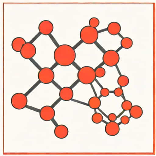
Understanding the structure of the hemoglobin molecule is essential for appreciating its critical role in transporting oxygen throughout the human body. Hemoglobin is a complex protein found in red blood cells, responsible for carrying oxygen from the lungs to tissues and returning carbon dioxide back to the lungs for exhalation. Its unique structure allows it to bind oxygen efficiently and release it where it is needed most, making it indispensable for survival. Schematic representations of hemoglobin provide a clear visual understanding of its composition, subunits, and binding sites, which can help students, researchers, and medical professionals grasp its functional mechanisms more easily. While hemoglobin is often discussed in textbooks with detailed 3D models, a simplified schematic drawing highlights the key structural elements and interactions that define its functionality.
Basic Structure of Hemoglobin
Hemoglobin is a tetrameric protein, meaning it is composed of four polypeptide chains. In adult humans, these chains are typically two alpha (α) chains and two beta (β) chains. Each of these subunits contains a heme group, which is a prosthetic group responsible for oxygen binding. The heme group includes an iron ion (Fe²⁺) at its center, which directly interacts with oxygen molecules. The quaternary structure of hemoglobin allows cooperative binding, meaning that when one heme group binds to oxygen, it increases the affinity of the remaining heme groups for oxygen. This property is crucial for efficient oxygen transport in varying oxygen concentrations throughout the body.
Polypeptide Chains and Heme Groups
- Each α and β chain consists of approximately 140-150 amino acids.
- Each chain contains a heme group that binds oxygen via the iron ion at its center.
- The arrangement of the four chains forms a roughly tetrahedral structure, providing stability and functional efficiency.
Schematic Representation of Hemoglobin
A schematic drawing of the hemoglobin molecule simplifies the complex 3D structure into an easy-to-understand diagram. The main elements to represent include the four polypeptide chains, the heme groups within each chain, and the iron ions. In diagrams, α-chains and β-chains are often shown in different colors for clarity, and heme groups are depicted as circular or hexagonal structures with the central iron ion marked. Oxygen molecules can be illustrated as small dots or spheres bound to the iron ions. Schematic drawings also often indicate the cooperative binding mechanism, showing how oxygen binding to one subunit influences the others.
Key Features to Include in a Schematic
- Four polypeptide chains (two α, two β), often color-coded.
- Heme groups within each chain, with central iron ions.
- Oxygen binding sites, indicating how oxygen attaches to iron.
- Connections between chains to represent the quaternary structure.
Oxygen Binding and Release
The function of hemoglobin is intimately tied to its structure. The iron ion in each heme group can reversibly bind one oxygen molecule. When hemoglobin reaches the oxygen-rich environment of the lungs, oxygen binds to the iron ions in each heme, converting hemoglobin to its oxygenated form, called oxyhemoglobin. As blood circulates to tissues with lower oxygen concentrations, hemoglobin releases oxygen where it is needed. The cooperative binding mechanism ensures that hemoglobin can efficiently pick up oxygen in the lungs and release it in tissues. Schematic diagrams often illustrate this dynamic process by showing hemoglobin in both its oxygenated and deoxygenated forms.
Cooperative Binding Explained
- Binding of the first oxygen molecule changes the shape of hemoglobin, making it easier for subsequent oxygen molecules to bind.
- This conformational change is represented in schematics by slight shifts in the relative positions of the subunits.
- Deoxygenation reverses the conformational change, facilitating oxygen release in tissues.
Applications of Hemoglobin Schematics
Schematic diagrams of hemoglobin are widely used in educational and clinical settings. They help students learn about protein structure and function, illustrate the importance of iron in oxygen transport, and demonstrate how mutations in hemoglobin subunits can lead to disorders such as sickle cell anemia or thalassemia. In research, schematics provide a simplified visual framework for discussing hemoglobin interactions with drugs, oxygen affinity, and allosteric regulators like 2,3-bisphosphoglycerate (2,3-BPG). In clinical contexts, understanding hemoglobin schematics is essential for interpreting blood tests, diagnosing anemia, and understanding oxygen delivery in critical care settings.
Common Disorders Related to Hemoglobin Structure
- Sickle Cell Anemia Caused by a mutation in the β-chain that leads to abnormal hemoglobin structure and red blood cell deformation.
- Thalassemia Resulting from defective synthesis of α or β chains, leading to imbalanced hemoglobin production.
- Methemoglobinemia Occurs when iron in the heme group is oxidized and cannot bind oxygen effectively.
How to Draw Hemoglobin Schematic Step-by-Step
Creating a clear schematic of hemoglobin involves several steps. Start by drawing four ovals or circles to represent the α and β chains. Use different colors or labels for clarity. Inside each chain, draw a heme group, often as a hexagon or circle, and mark the central iron ion. Connect the chains to illustrate the quaternary structure, showing how the four subunits interact. Optionally, indicate oxygen molecules bound to the iron ions and arrows to show cooperative binding. Adding these details creates a comprehensive schematic that communicates both structure and function effectively.
Step-by-Step Tips
- Draw four separate chains and label them α and β.
- Insert heme groups in each chain and mark the iron ion.
- Connect the chains to form the tetrameric structure.
- Show oxygen molecules bound to iron ions.
- Indicate conformational changes to illustrate cooperative binding.
In summary, drawing a schematic of the hemoglobin molecule provides an accessible way to understand one of the most important proteins in the human body. Hemoglobin’s tetrameric structure, with four polypeptide chains and heme groups containing iron ions, enables efficient oxygen transport and release. Schematics highlight key features such as oxygen binding sites, cooperative binding, and the interactions between subunits, making complex molecular details more understandable. Understanding hemoglobin schematically also aids in education, research, and clinical practice, providing insights into both normal physiological function and the effects of genetic disorders on oxygen transport. By following step-by-step schematic drawing methods, one can capture the essence of hemoglobin’s structure and function, making it a powerful tool for learning and communication in biology and medicine.