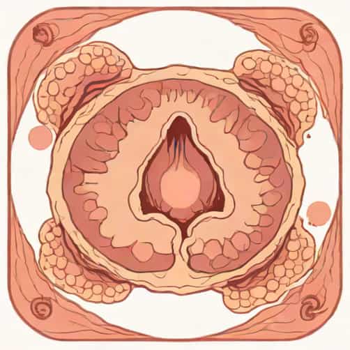
Cystadenoma serous of the ovary is a common type of benign ovarian tumor that is frequently encountered in gynecologic pathology. Understanding its pathology is crucial for accurate diagnosis, proper management, and effective patient counseling. Serous cystadenomas are typically characterized by their cystic nature and serous fluid content, and they are most often seen in women of reproductive age, although they can occur at any stage of life. Pathology outlines for cystadenoma serous ovary provide detailed guidance for pathologists and clinicians to identify histological features, differentiate from other ovarian tumors, and assess clinical implications.
Overview of Serous Cystadenoma
Serous cystadenomas are epithelial ovarian tumors arising from the ovarian surface epithelium. They are part of the broader category of ovarian cystadenomas, which also includes mucinous cystadenomas. Serous cystadenomas are typically unilateral, although bilateral cases may occur in a small percentage of patients. They usually present as unilocular or multilocular cysts with smooth inner linings and contain clear, serous fluid. These tumors are generally benign, with low potential for malignant transformation, but accurate histopathological assessment is essential to exclude borderline or malignant forms.
Clinical Presentation
Patients with serous cystadenomas often present with nonspecific symptoms, which may include abdominal distension, pelvic pain, or pressure symptoms due to the size of the cyst. Some cases are discovered incidentally during imaging for unrelated conditions. Physical examination may reveal a palpable adnexal mass, and imaging techniques such as ultrasound or MRI are typically used to assess the size, morphology, and cystic nature of the tumor. Serous cystadenomas are usually asymptomatic when small, and symptoms tend to correlate with tumor size.
Pathology and Histological Features
Histologically, serous cystadenomas are lined by a single layer of columnar or cuboidal epithelial cells that resemble fallopian tube epithelium. The epithelial lining may contain ciliated and non-ciliated cells, and there is often a fibrous stroma supporting the cyst. Key features in pathology outlines include the absence of significant cellular atypia, stromal invasion, or papillary structures, which helps differentiate benign cystadenomas from borderline or malignant serous tumors.
Gross Examination
On gross examination, serous cystadenomas are usually smooth-walled, thin-walled cysts that may be unilocular or multilocular. The cysts contain clear or straw-colored fluid and are typically well-circumscribed. Bilateral involvement is uncommon, and the tumors rarely exceed 10-15 cm in diameter. Gross features are important in pathology outlines to guide tissue sampling for histological examination and to identify any areas suspicious for borderline or malignant transformation.
Microscopic Characteristics
- Single layer of uniform epithelial cells lining the cyst.
- Presence of ciliated and non-ciliated columnar or cuboidal cells.
- Minimal to no cytologic atypia.
- Fibrous stromal support without invasion into surrounding ovarian tissue.
- Absence of complex papillary structures, which are seen in serous borderline tumors.
Immunohistochemical and Molecular Findings
Immunohistochemistry can aid in differentiating serous cystadenomas from other ovarian neoplasms. Typical markers include strong expression of cytokeratin, PAX8, and WT1, which are characteristic of serous differentiation. Molecular studies often reveal fewer genetic alterations in benign serous cystadenomas compared to borderline or malignant tumors. Mutations in TP53, KRAS, or BRAF are usually absent in benign cystadenomas, supporting their non-aggressive nature.
Pathology Outlines Recommendations
Pathology outlines emphasize a systematic approach to diagnosis, which includes
- Gross inspection of the cyst for size, laterality, and fluid characteristics.
- Comprehensive sampling of the cyst wall to assess epithelial lining.
- Microscopic evaluation for cellular atypia, ciliation, and stromal features.
- Use of immunohistochemical stains to confirm serous differentiation.
- Documentation of findings in a standardized pathology report for clinical correlation.
Differential Diagnosis
Serous cystadenomas must be differentiated from other ovarian cystic lesions, including mucinous cystadenomas, endometriomas, cystic teratomas, and borderline or malignant serous tumors. Mucinous cystadenomas contain mucin rather than serous fluid and often have more complex septations. Endometriomas are associated with hemorrhagic content and endometrial stroma. Careful histological evaluation and immunohistochemical studies are essential to accurately classify the lesion and guide clinical management.
Clinical Management
The management of serous cystadenomas of the ovary typically involves surgical removal, especially if the cyst is symptomatic, large, or increasing in size. Laparoscopic cystectomy is commonly performed, and oophorectomy may be indicated in selected cases. Complete excision usually results in an excellent prognosis, with minimal risk of recurrence. Follow-up may include periodic imaging to ensure no new cysts develop, especially in patients with bilateral involvement or a history of ovarian cysts.
Prognosis and Follow-Up
Patients with benign serous cystadenomas generally have an excellent prognosis. Recurrence is rare after complete excision. Pathology outlines highlight the importance of distinguishing benign cystadenomas from borderline and malignant tumors to avoid overtreatment or unnecessary aggressive surgery. Counseling patients about the benign nature of the tumor and the low risk of malignancy is an important component of postoperative care.
Educational Value of Pathology Outlines
Pathology outlines for cystadenoma serous ovary serve as a critical resource for medical students, residents, and practicing pathologists. They provide concise, structured information on the histopathological, gross, and clinical features of the tumor. By following these outlines, healthcare professionals can ensure accurate diagnosis, appropriate management, and effective communication with patients and colleagues.
Serous cystadenoma of the ovary is a common, benign epithelial tumor that requires careful pathological evaluation to ensure accurate diagnosis and appropriate management. Pathology outlines provide a systematic framework for assessing gross and microscopic features, differentiating the tumor from other ovarian lesions, and guiding surgical and clinical decisions. With proper identification and treatment, patients can expect excellent outcomes, reinforcing the importance of thorough understanding and adherence to pathology guidelines in gynecologic practice.