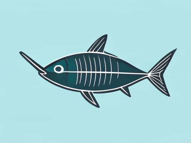
When studying marine life, one of the most interesting groups to explore is the class of cartilaginous fish, scientifically known as Chondrichthyes. These animals include sharks, rays, skates, and chimaeras, all sharing a similar body structure that can be outlined even without an actual drawing. While a diagram of a cartilaginous fish typically highlights the major anatomical features such as fins, gills, muscle structure, and internal organs, it is just as useful to understand these parts through explanation. Knowing how each feature works together not only helps students and enthusiasts visualize a cartilaginous fish diagram mentally, but also builds a deeper appreciation for how these ancient species have survived for millions of years in diverse marine environments.
General Body Structure
A cartilaginous fish diagram usually begins with the streamlined body shape, which helps these animals move efficiently through water. The body is covered in dermal denticles instead of scales, giving the skin a rough texture that reduces drag. Beneath the tough exterior is a skeleton made entirely of cartilage, which provides strength and flexibility while weighing less than bone.
Head Region
At the front of the body is the head, which contains sensory organs crucial for survival. Even without viewing a diagram of a cartilaginous fish, it is helpful to picture the placement of the eyes on either side of the head, allowing for wide peripheral vision. The mouth is located below the snout, a position that supports bottom feeding as well as prey capture in open water.
- Eyes adapted to low light conditions
- Nostrils for detecting scent trails
- Ampullae of Lorenzini for sensing electrical signals
These sensory systems show why the head dominates attention in many anatomical illustrations. The ampullae of Lorenzini, tiny pores clustered near the snout, are rarely shown in basic diagrams, yet they are essential features that allow cartilaginous fish to detect movement and prey hidden in sand or deeper waters.
Fins and Mobility
Another significant part labeled in a typical diagram of a cartilaginous fish is the fin arrangement. Movement relies on the coordinated action of several fins, each serving a different purpose. The pectoral fins, located near the head, act like wings, giving lift and steering ability. Meanwhile, the dorsal fins provide stability, preventing the body from rolling side to side.
Types of Fins
- Pectoral fins steering and lift
- Dorsal fins balance and defense
- Pelvic fins stability during swimming
- Caudal fin propulsion and acceleration
In most diagrams, the caudal fin at the tail is drawn with an asymmetrical shape, known as a heterocercal tail. This uneven design generates strong thrust, helping sharks and rays move powerfully with fewer strokes. The placement and size of each fin vary among cartilaginous species, but the general layout remains consistent across the group.
Internal Anatomy Overview
While external features are usually easy to identify in a diagram of a cartilaginous fish, internal structures require more explanation. These fish have a muscular body with a large liver that aids in buoyancy because they lack a swim bladder. The digestive system includes a spiral valve intestine, a unique feature that increases the surface area for nutrient absorption without requiring long intestinal length.
Key Internal Structures
- Heart with two chambers for circulating blood
- Gills for extracting oxygen from water
- Liver for buoyancy and fat storage
- Spiral valve intestine for digestion
The gills, located behind the head, are arranged in multiple slits rather than a single operculum like those found in bony fish. In most cartilaginous fish, five to seven gill slits appear on each side of the body. This design is often clearly labeled in anatomical outlines, reinforcing how respiration works without involving a swim bladder.
Respiratory System Details
Breathing mechanisms in these fish depend on constant water flow over the gills. A physical diagram of a cartilaginous fish usually marks the spiracles behind the eyes, openings that allow some species to breathe while resting on the sea floor. Rays and skates rely heavily on spiracles because they often bury themselves in sand.
How Gills Function
Water enters through the mouth or spiracles and passes over the gill filaments where oxygen exchange occurs. The structure of these filaments increases surface area, allowing efficient absorption of oxygen even in low-oxygen environments. This respiratory efficiency is part of what makes cartilaginous fish powerful swimmers and effective predators.
Reproductive Anatomy
A diagram of a cartilaginous fish sometimes includes reproductive organs, especially for instructional purposes. Male cartilaginous fish have claspers located near the pelvic fins, which are used during internal fertilization. Females may be oviparous, ovoviviparous, or viviparous depending on the species.
Reproduction Methods
- Oviparous eggs laid outside the body
- Ovoviviparous eggs hatch internally before live birth
- Viviparous embryos nourished internally via connection
Internal fertilization gives offspring a better chance of survival compared to external spawning. Even in the absence of a visual diagram, understanding the reproductive system helps explain why cartilaginous fish populations grow differently compared to bony fish.
Sensory System Highlight
One of the most fascinating elements featured in educational diagrams is the advanced sensory system. Besides vision and smell, cartilaginous fish possess lateral lines that detect vibrations and water movement, helping them sense nearby animals. This line runs along the body from head to tail and is often outlined in diagrams to show how it is integrated into the fish’s nervous system.
Electroreception and Adaptation
The ability to detect electrical pulses is vital for hunting and navigation in dark or murky waters. Each pore of the ampullae of Lorenzini connects to nerve fibers, making the head region extremely sensitive. This specialized adaptation is one reason sharks can locate prey hidden beneath sand or even detect weak electrical activity emitted by struggling fish.
Why Understanding the Diagram Matters
Learning the anatomy of cartilaginous fish through descriptive detail can replace the need for a physical diagram while still offering clarity. Knowing where structures are located and how they function helps students connect behavior, physiology, and evolution. The streamlined body, flexible cartilage skeleton, and sensory organs all work together to support survival strategies that have remained effective for millions of years.
Connecting Anatomy to Ecology
With a firm mental image of how the features align, it becomes easier to understand how sharks and rays fit into marine food webs. Their mobility, sensory perception, and efficient digestion allow them to thrive as apex predators or bottom feeders depending on species. Understanding anatomy builds the foundation for ecological awareness, conservation, and deeper scientific study.
A diagram of a cartilaginous fish typically labels external fins, gill slits, internal organs, and sensory systems, but these details can be interpreted just as clearly through descriptive explanations. By examining each body region and understanding how it contributes to survival, it becomes easier to visualize the organism without needing a picture. Studying the anatomy prepares learners to explore species diversity, habitat adaptation, and biological evolution, making the subject both accessible and rewarding for anyone curious about marine life.