Obtaining clear and precise images of the foot is crucial in diagnosing fractures, dislocations, arthritis, and other abnormalities. One of the specialized views used in radiography is the oblique view of the foot, which provides a unique perspective that cannot be captured through standard anterior-posterior or lateral views. Understanding the proper positioning for an X-ray foot oblique view is essential for radiologic technologists, medical students, and healthcare professionals to ensure accurate imaging and proper diagnosis of foot conditions.
Understanding the Oblique View
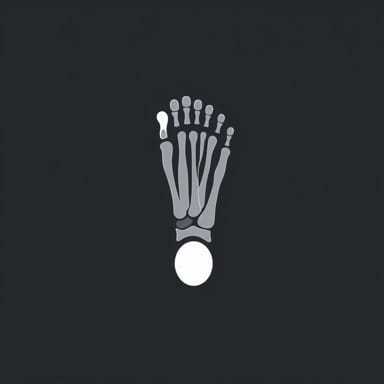
The oblique view of the foot is a radiographic technique that captures the foot at an angle, typically around 30 to 45 degrees from the plane of the table or detector. This angled perspective allows better visualization of the metatarsals, tarsal bones, and joints, which may overlap in standard AP (anteroposterior) or lateral views. By using the oblique view, clinicians can detect subtle fractures, joint space abnormalities, and bone lesions that may not be apparent in other projections.
Purpose of the Oblique View
The primary purpose of the X-ray foot oblique view is to provide a more detailed examination of the foot’s skeletal structure. Some specific reasons for performing this view include
- Identifying fractures in the metatarsals, cuboid, or cuneiform bones.
- Evaluating joint alignment and spacing in cases of arthritis or trauma.
- Detecting foreign bodies or bone spurs that may affect mobility.
- Assessing post-operative healing after foot surgery.
- Providing comprehensive imaging for patients with foot pain or deformities.
Patient Preparation
Proper patient preparation is crucial for obtaining a clear oblique X-ray of the foot. Preparation steps include
- Explaining the procedure to the patient to ensure cooperation.
- Removing any footwear, socks, or jewelry that may interfere with imaging.
- Positioning the patient comfortably, either seated or lying on the examination table, depending on equipment setup.
- Ensuring the foot is clean and dry to prevent artifacts on the X-ray film.
Positioning for X-Ray Foot Oblique View
Accurate positioning is essential to capture the oblique view effectively. The foot must be rotated and aligned correctly to highlight specific bones and joints.
Steps for Proper Positioning
- Patient PositionThe patient may be seated with the leg extended or lying supine on the table. The knee can be slightly flexed to relax the foot muscles.
- Foot PlacementThe foot is placed flat on the image receptor or table surface, depending on the equipment. The toes should be pointing upward or slightly dorsiflexed.
- RotationRotate the foot medially or laterally, typically at an angle of 30 to 45 degrees. This angle allows the metatarsals and tarsal bones to be separated visually, reducing overlap.
- CenteringThe central ray should be directed to the base of the third metatarsal, ensuring that the entire foot is included in the image.
- StabilizationEnsure the foot remains still during the exposure to prevent blurring. Support devices or foam wedges may be used to maintain the position.
Radiation Safety Considerations
Maintaining safety for both the patient and radiology personnel is critical. Steps include
- Using lead aprons or shields to protect other parts of the body from unnecessary exposure.
- Confirming the X-ray beam is collimated to the area of interest, minimizing radiation dose.
- Following ALARA principles (As Low As Reasonably Achievable) to reduce radiation exposure.
- Ensuring proper alignment of the X-ray equipment to prevent repeat exposures.
Common Variations of the Foot Oblique View
Different oblique angles may be used depending on the clinical indication. Variations include
- Medial ObliqueThe foot is rotated inward toward the midline of the body, typically used to visualize the lateral tarsal bones and metatarsals.
- Lateral ObliqueThe foot is rotated outward, which can highlight the medial structures, including the navicular and first cuneiform bones.
- Weight-Bearing ObliqueIn some cases, the patient may be asked to stand on the affected foot to assess joint spaces and arch stability under load.
Interpretation of Oblique Foot X-Ray
Once the image is captured, radiologists or orthopedic specialists interpret the oblique view in conjunction with other foot X-rays. Key structures evaluated include
- Metatarsals for fractures, dislocations, or alignment issues.
- Tarsal bones such as cuboid, navicular, and cuneiforms for abnormalities.
- Joint spaces, including the metatarsophalangeal and intertarsal joints, to detect arthritis or injury.
- Soft tissue shadows to identify swelling, foreign bodies, or masses.
- Signs of bone healing or post-surgical changes if applicable.
Common Clinical Indications
The X-ray foot oblique view is commonly requested in the following situations
- Acute trauma, such as suspected metatarsal or tarsal fractures.
- Chronic foot pain with unclear etiology after standard AP and lateral views.
- Evaluation of foot deformities, such as bunions or claw toes.
- Pre- and post-operative assessment for orthopedic procedures.
- Monitoring bone healing in pediatric or adult patients with previous fractures.
Tips for Successful Imaging
To ensure high-quality oblique foot X-rays, consider these tips
- Ensure the foot is properly dorsiflexed to prevent overlapping of the toes and metatarsals.
- Use appropriate exposure settings to visualize both cortical and trabecular bone.
- Double-check foot rotation and central ray alignment before exposure.
- Maintain patient comfort to reduce movement during the procedure.
- Document the view (medial or lateral oblique) and any clinical indications for future reference.
The X-ray foot oblique view is an essential imaging technique that provides a detailed perspective of the foot’s skeletal structures, especially the metatarsals and tarsal bones. Proper positioning, including rotation, foot placement, and central ray alignment, is crucial to obtain clear, diagnostic-quality images. This view helps detect fractures, joint abnormalities, and other foot pathologies that may not be visible in standard AP or lateral projections. Radiologists and orthopedic specialists rely on accurate oblique foot images to make informed clinical decisions, guide treatment plans, and monitor patient recovery. By understanding the positioning, purpose, and interpretation of the foot oblique view, healthcare professionals can ensure accurate diagnosis, improved patient care, and optimal outcomes in foot imaging.