Fractures involving the distal radius and ulnar styloid are common injuries, especially among individuals who experience falls onto an outstretched hand. These types of fractures can range from simple, non-displaced breaks to complex, comminuted injuries that affect wrist function and stability. Understanding the anatomy, mechanism of injury, symptoms, diagnosis, and treatment options is essential for effective management and optimal recovery. Early intervention and proper rehabilitation can significantly impact the patient’s ability to regain full wrist motion and strength.
Anatomy of the Distal Radius and Ulnar Styloid
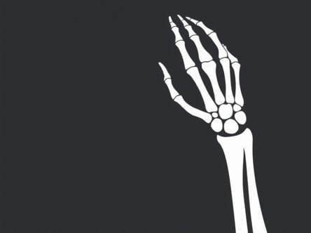
The distal radius is the end portion of the radius bone near the wrist joint. It plays a crucial role in wrist articulation, supporting the hand and enabling a wide range of motion. The ulnar styloid is a bony prominence at the distal end of the ulna, positioned on the medial side of the wrist. It serves as an attachment point for the triangular fibrocartilage complex (TFCC), which stabilizes the distal radioulnar joint (DRUJ) and facilitates smooth wrist movements.
Mechanism of Injury
Ulnar styloid and distal radius fractures commonly occur due to trauma, typically from a fall onto an outstretched hand. High-energy impacts, such as those from motor vehicle accidents or sports injuries, can also result in these fractures. The severity of the fracture depends on the force and angle of impact, bone quality, and patient age. Older adults, especially those with osteoporosis, are more prone to distal radius fractures from relatively minor falls.
Types of Distal Radius Fractures
Distal radius fractures can be classified based on fracture pattern, displacement, and involvement of the joint surface
- Colles’ fractureA common type with dorsal displacement of the distal fragment.
- Smith’s fractureInvolves volar displacement of the distal fragment.
- Intra-articular fractureThe fracture extends into the wrist joint, potentially affecting joint stability and motion.
- Extra-articular fractureThe break does not involve the joint surface, often simpler to manage.
Ulnar Styloid Fractures
Fractures of the ulnar styloid often occur alongside distal radius fractures. They can be classified as
- Tip fracturesInvolve the distal tip of the styloid and typically do not affect DRUJ stability.
- Base fracturesOccur at the base of the styloid and may compromise TFCC attachment, affecting wrist stability.
Ulnar styloid fractures may sometimes be asymptomatic but can lead to persistent wrist pain or instability if associated with DRUJ injury.
Symptoms of Distal Radius and Ulnar Styloid Fractures
Patients with these fractures typically present with
- Immediate pain and swelling around the wrist.
- Visible deformity, such as a dinner fork appearance in Colles’ fractures.
- Bruising and tenderness on palpation of the distal radius or ulnar styloid.
- Reduced range of motion and difficulty gripping objects.
- Numbness or tingling in the hand if nerve compression occurs.
Diagnosis
Accurate diagnosis relies on a combination of physical examination and imaging studies
- X-raysStandard anteroposterior (AP) and lateral views are used to assess fracture pattern, displacement, and involvement of the joint surface.
- CT scanMay be indicated for complex intra-articular fractures to guide surgical planning.
- MRIRarely required but can evaluate soft tissue involvement, particularly TFCC injuries.
Treatment Options
Management depends on fracture type, displacement, patient age, and functional demands. Treatment can be broadly divided into non-surgical and surgical approaches.
Non-Surgical Management
Non-displaced or minimally displaced fractures may be treated conservatively with
- ImmobilizationUse of a cast or splint for 4-6 weeks to allow bone healing.
- Elevation and iceReduce swelling and pain.
- Pain managementAnalgesics and anti-inflammatory medications.
- Physical therapyInitiated after cast removal to restore motion, strength, and function.
Surgical Management
Surgical intervention is indicated for
- Displaced fractures with significant angulation or shortening.
- Intra-articular fractures compromising joint congruency.
- Fractures associated with DRUJ instability.
- Failed conservative treatment with persistent displacement or pain.
Surgical options include
- Open reduction and internal fixation (ORIF)Plates and screws are used to realign the distal radius fragments.
- External fixationApplied for comminuted fractures or severe soft tissue injury.
- Ulnar styloid fixationTypically reserved for base fractures affecting DRUJ stability.
Rehabilitation and Recovery
Recovery depends on fracture severity, treatment method, and patient compliance with rehabilitation protocols. Physical therapy focuses on
- Restoring wrist range of motion and flexibility.
- Strengthening forearm and hand muscles.
- Improving grip strength and dexterity.
- Reducing stiffness and swelling through controlled exercises and modalities.
Most patients regain functional wrist motion within 3-6 months, though full recovery may take longer for complex fractures.
Complications
Potential complications of distal radius and ulnar styloid fractures include
- Malunion or improper bone healing leading to deformity.
- DRUJ instability, particularly with ulnar styloid base fractures.
- Chronic pain or post-traumatic arthritis.
- Nerve injury causing numbness or tingling in the hand.
- Reduced range of motion or grip strength if rehabilitation is inadequate.
Fractures of the distal radius and ulnar styloid are common wrist injuries that require careful evaluation and appropriate management. Understanding the anatomy, fracture types, and mechanisms of injury is essential for determining the best treatment approach. While non-surgical management is effective for many stable fractures, surgical intervention may be necessary for displaced or unstable injuries. Comprehensive rehabilitation is crucial to restore function, prevent complications, and ensure optimal recovery. Early diagnosis, proper treatment, and patient adherence to rehabilitation protocols can significantly improve outcomes and reduce long-term disability associated with these fractures.