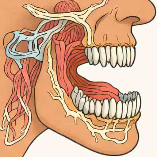
The muscles of mastication play a crucial role in the basic yet essential act of chewing. These muscles are responsible for the movements of the jaw, allowing us to bite, grind, and manipulate food in preparation for swallowing and digestion. The nerve supply of the muscles of mastication is primarily derived from the mandibular division of the trigeminal nerve, which is the fifth cranial nerve. Understanding the anatomy and neural control of these muscles is not only important for students of medicine and dentistry but also for clinicians dealing with disorders of the jaw, temporomandibular joint (TMJ), or facial pain syndromes. This topic provides valuable insight into how the nervous system integrates motor control with coordinated muscle activity to perform one of the most vital functions of daily life.
Overview of the Muscles of Mastication
The term muscles of mastication refers to a group of four primary muscles that move the mandible (lower jaw) during chewing. These include
- Masseter
- Temporalis
- Medial pterygoid
- Lateral pterygoid
All of these muscles are paired, meaning there is one on each side of the face. They work together to elevate, depress, protrude, retract, and move the mandible side to side. While their functions differ slightly, their common feature is their nerve supply, which comes from the motor root of the mandibular nerve the third division of the trigeminal nerve (cranial nerve V).
The Trigeminal Nerve Source of Motor Innervation
The trigeminal nerve is the largest of the cranial nerves and has both sensory and motor components. It divides into three major branches
- Ophthalmic division (V1)
- Maxillary division (V2)
- Mandibular division (V3)
Among these, only the mandibular division (V3) carries motor fibers, which supply the muscles of mastication and a few other smaller muscles in the head. The motor root of the trigeminal nerve originates in the motor nucleus of the trigeminal nerve located in the pons of the brainstem. From there, the fibers travel with the sensory portion of V3 to reach their target muscles in the lower face.
Mandibular Nerve and Its Motor Branches
The mandibular nerve exits the skull through the foramen ovale and immediately divides into anterior and posterior divisions. The motor branches supplying the muscles of mastication arise mainly from the anterior division. However, there are some exceptions, such as the nerve to the mylohyoid, which comes from the posterior division. Below is an overview of how each muscle receives its nerve supply.
1. Masseter Muscle
The masseter is a thick, rectangular muscle located at the angle of the jaw. It elevates the mandible, contributing to the closing of the mouth and the clenching of teeth. The nerve supply to the masseter comes from themasseteric nerve, a branch of the anterior division of the mandibular nerve. This nerve passes through the mandibular notch to enter the deep surface of the muscle.
2. Temporalis Muscle
The temporalis is a broad, fan-shaped muscle situated on the side of the skull. Its fibers converge downward to insert on the coronoid process of the mandible. The muscle assists in elevating and retracting the mandible. The nerve supply to the temporalis comes from thedeep temporal nerves, typically two or three small branches from the anterior division of the mandibular nerve. These nerves enter the deep surface of the muscle to provide motor innervation.
3. Medial Pterygoid Muscle
The medial pterygoid is located deep in the face, running parallel to the masseter but on the inner surface of the mandible. It elevates the mandible and helps in side-to-side movements during chewing. Thenerve to the medial pterygoidarises directly from the main trunk of the mandibular nerve before the division into anterior and posterior branches. Interestingly, this same nerve also sends small branches to the tensor tympani and tensor veli palatini muscles in the middle ear and soft palate, respectively.
4. Lateral Pterygoid Muscle
The lateral pterygoid is the only muscle of mastication primarily responsible for opening the mouth by depressing and protruding the mandible. It also assists in lateral jaw movements. It receives its nerve supply from thelateral pterygoid nerve, another branch of the anterior division of the mandibular nerve. This nerve typically divides into two branches that enter both the superior and inferior heads of the muscle.
Accessory Muscles Involved in Mastication
While the four main muscles perform the majority of chewing movements, several accessory muscles assist in the process. These include the buccinator, mylohyoid, and digastric muscles. Though they are not strictly classified as muscles of mastication, their functions complement chewing and swallowing.
- BuccinatorKeeps food between the teeth by compressing the cheeks during chewing. It is supplied by the facial nerve, not the trigeminal nerve.
- MylohyoidHelps elevate the floor of the mouth during swallowing. It receives its nerve supply from the mylohyoid branch of the inferior alveolar nerve, a branch of the mandibular nerve.
- Anterior belly of digastricAssists in depressing the mandible and is supplied by the same mylohyoid branch.
Functional Coordination of Nerve Supply
The process of mastication involves precise coordination between the muscles and their nerve supply. The motor neurons in the trigeminal motor nucleus are controlled by higher brain centers such as the cerebral cortex and cerebellum, ensuring smooth and rhythmic jaw movements. Sensory feedback from the teeth, temporomandibular joint, and muscles helps regulate the force and timing of contractions. This integration allows for efficient grinding of food and protection against overstrain or injury to the jaw.
Clinical Significance
Damage to the mandibular division of the trigeminal nerve or its branches can lead to serious functional deficits. Common clinical implications include
- Weakness of masticationDifficulty in chewing due to paralysis of the jaw muscles.
- Deviation of the jawWhen the patient opens the mouth, the mandible deviates toward the paralyzed side because the lateral pterygoid muscle on that side fails to contract properly.
- Loss of sensationSince the mandibular nerve also carries sensory fibers, injury may result in numbness in the lower face, teeth, or tongue.
- Temporomandibular joint dysfunctionImproper muscle coordination can lead to TMJ pain, clicking, or restricted movement.
Electromyography (EMG) and nerve conduction studies are often used to evaluate the function of these nerves in clinical diagnosis. In some cases, dental procedures, fractures, or surgical interventions near the mandibular region can inadvertently damage the motor branches, requiring rehabilitation and therapy to restore function.
Summary of Nerve Supply
To summarize, the muscles of mastication receive their motor innervation from the mandibular division of the trigeminal nerve (cranial nerve V3), specifically through the following branches
- Masseter Masseteric nerve
- Temporalis Deep temporal nerves
- Medial pterygoid Nerve to medial pterygoid
- Lateral pterygoid Nerve to lateral pterygoid
This consistent pattern of innervation emphasizes the close functional relationship between the trigeminal nerve and the masticatory apparatus.
The nerve supply of the muscles of mastication is a prime example of how anatomy and physiology work in harmony to support one of the most essential human activities chewing. All four primary muscles receive motor input from the mandibular division of the trigeminal nerve, ensuring precise and coordinated jaw movements. Any disruption in this neural pathway can significantly affect oral function, making this anatomical relationship vital for clinicians, dentists, and surgeons to understand. Through its intricate network of nerves and muscles, the system of mastication highlights the remarkable design and efficiency of the human body.