The muscles of mastication are essential for chewing and grinding food, playing a critical role in the process of digestion and overall oral function. Proper innervation of these muscles ensures coordinated movement and strength required for effective mastication. Understanding the innervation of the muscles of mastication is fundamental for students of anatomy, dentistry, and medicine, as well as for professionals involved in maxillofacial surgery or neurology. This topic provides a comprehensive overview of the nerve supply to the muscles of mastication, their functions, and clinical significance, making it easier for readers to grasp the complexities of cranial nerve anatomy.
Overview of the Muscles of Mastication
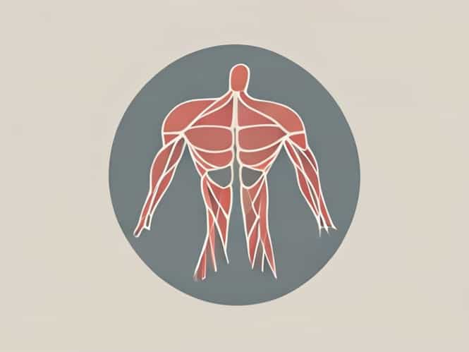
The muscles of mastication are primarily responsible for the movement of the mandible during chewing. These muscles include the masseter, temporalis, medial pterygoid, and lateral pterygoid. Each of these muscles has a specific role in elevating, depressing, protruding, or moving the mandible laterally. Proper function of these muscles depends on accurate and coordinated innervation by cranial nerves, particularly branches of the trigeminal nerve. Dysfunction in innervation can result in difficulty chewing, jaw weakness, or abnormal movements.
Major Muscles of Mastication
- MasseterElevates the mandible, allowing for forceful closure of the jaw.
- TemporalisElevates and retracts the mandible, assisting in chewing and biting.
- Medial PterygoidElevates the mandible and contributes to side-to-side movements.
- Lateral PterygoidDepresses and protrudes the mandible, enabling lateral movements and opening the jaw.
Innervation of the Muscles of Mastication
The primary nerve responsible for innervating the muscles of mastication is the mandibular division of the trigeminal nerve, also known as cranial nerve V3. The trigeminal nerve is the fifth cranial nerve and has three major branches ophthalmic (V1), maxillary (V2), and mandibular (V3). Only the mandibular branch provides motor fibers to the muscles of mastication, along with some sensory fibers for the lower face, oral cavity, and mandibular teeth.
Mandibular Nerve (V3)
The mandibular nerve exits the skull through the foramen ovale and immediately divides into anterior and posterior branches. The anterior division is mainly motor, while the posterior division is primarily sensory. Both branches contribute to the precise control of the muscles of mastication. Each muscle receives its motor supply through specific branches of the mandibular nerve, ensuring coordinated jaw movements during chewing.
Specific Innervation of Each Muscle
Masseter Muscle
The masseter is innervated by the masseteric nerve, which is a branch of the anterior division of the mandibular nerve. This nerve travels through the mandibular notch to reach the masseter muscle. Innervation allows the masseter to generate strong forces required for closing the jaw, making it one of the most powerful muscles in the body relative to its size.
Temporalis Muscle
The temporalis muscle receives its motor fibers from the deep temporal nerves, also arising from the anterior division of the mandibular nerve. These nerves enter the temporalis muscle at multiple points, enabling fine control over elevation and retraction of the mandible. The coordinated action of the temporalis is critical for biting and maintaining occlusion during chewing.
Medial Pterygoid Muscle
The medial pterygoid is innervated by the nerve to the medial pterygoid, which is a small branch of the mandibular nerve. Interestingly, this nerve also provides fibers to the tensor veli palatini and tensor tympani muscles, highlighting the close anatomical relationships in the region. The medial pterygoid works with the masseter to elevate the mandible and assists in grinding movements during mastication.
Lateral Pterygoid Muscle
The lateral pterygoid muscle receives its innervation from the nerve to the lateral pterygoid, which comes from the anterior division of V3. The lateral pterygoid has two heads superior and inferior. Both heads are involved in protruding the mandible, depressing the jaw, and producing side-to-side movements necessary for proper grinding of food. Dysfunction in this nerve can lead to difficulty opening the mouth or asymmetrical jaw movements.
Additional Muscles and Accessory Innervation
While the primary muscles of mastication are directly innervated by branches of the mandibular nerve, there are accessory muscles, such as the tensor veli palatini, that indirectly support mastication by stabilizing the jaw and soft palate. These muscles also receive fibers from V3, demonstrating the multifunctional role of the mandibular nerve in coordinating oral and cranial functions. Proper innervation ensures smooth and controlled movements of the jaw during speech, chewing, and swallowing.
Clinical Significance
Understanding the innervation of the muscles of mastication is essential in clinical practice. Damage to the mandibular nerve can result in weakness or paralysis of the jaw muscles, leading to difficulty chewing, malocclusion, and atrophy of the affected muscles. Conditions such as trigeminal neuralgia, traumatic nerve injury, or surgical complications can disrupt innervation. Dentists, surgeons, and neurologists must consider the pathways of these nerves when planning procedures to avoid functional impairment.
Diagnostic Considerations
- Testing jaw strength and movement can help assess the function of the trigeminal nerve.
- Electromyography (EMG) can measure electrical activity in the muscles to detect nerve injury.
- Imaging studies, such as MRI, may be used to visualize nerve compression or lesions affecting V3.
- Knowledge of the specific branches allows targeted local anesthesia for dental or surgical procedures.
- Understanding nerve-muscle relationships is crucial for rehabilitation after nerve injury.
The innervation of the muscles of mastication is a complex yet highly organized system primarily controlled by the mandibular division of the trigeminal nerve (V3). Each muscle masseter, temporalis, medial pterygoid, and lateral pterygoid receives specific motor fibers that enable precise and coordinated jaw movements essential for chewing, speaking, and swallowing. Additional accessory muscles contribute to stabilization and overall oral function. Clinically, knowledge of these neural pathways is critical for diagnosing nerve injuries, planning surgeries, and administering effective anesthesia. A detailed understanding of the innervation of the muscles of mastication not only supports anatomical education but also enhances clinical practice in dentistry, maxillofacial surgery, and neurology.