The inferior cerebellar peduncle is a critical structure in the brain that plays a vital role in motor coordination, balance, and proprioception. It serves as one of the primary communication pathways between the cerebellum and the brainstem, carrying sensory information from the spinal cord and medulla to the cerebellum. Imaging this structure using MRI (Magnetic Resonance Imaging) provides essential insights for diagnosing neurological disorders, evaluating injuries, and planning surgical interventions. An understanding of the anatomy, function, and MRI characteristics of the inferior cerebellar peduncle is crucial for clinicians, radiologists, and neurologists when assessing patients with cerebellar or brainstem pathology.
Anatomy of the Inferior Cerebellar Peduncle
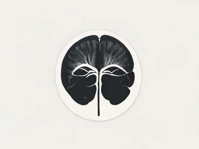
The inferior cerebellar peduncle, also known as the restiform body, is located in the posterior aspect of the brainstem. It connects the cerebellum to the medulla oblongata and carries afferent fibers primarily from the spinal cord and medulla to the cerebellar cortex. This peduncle is composed of multiple fiber tracts, including the dorsal spinocerebellar tract, cuneocerebellar tract, and olivocerebellar fibers. These fibers are crucial for transmitting information about body position, muscle tone, and reflexes, enabling the cerebellum to coordinate smooth and precise movements.
Structural Components
- Dorsal spinocerebellar tract transmits proprioceptive information from the lower limbs
- Cuneocerebellar tract carries sensory information from the upper limbs
- Olivocerebellar fibers convey signals from the inferior olivary nucleus to the cerebellum
- Vestibulocerebellar fibers integrate balance and spatial orientation information
Function of the Inferior Cerebellar Peduncle
The primary function of the inferior cerebellar peduncle is to relay sensory input from the spinal cord and medulla to the cerebellum for processing. By providing real-time feedback about limb position, muscle tone, and joint movements, it enables the cerebellum to coordinate voluntary and involuntary movements effectively. Additionally, the inferior cerebellar peduncle integrates vestibular signals, contributing to balance, posture, and equilibrium. Any damage or pathology affecting this peduncle can lead to significant deficits in coordination, tremors, ataxia, and imbalance.
Clinical Significance
- Essential for proprioception and motor coordination
- Contributes to the integration of sensory and vestibular information
- Lesions can result in ipsilateral cerebellar ataxia and limb incoordination
- Key structure evaluated in patients with stroke, demyelinating diseases, or tumors affecting the posterior fossa
MRI Imaging of the Inferior Cerebellar Peduncle
Magnetic Resonance Imaging (MRI) is the preferred modality for visualizing the inferior cerebellar peduncle due to its superior soft tissue contrast and multiplanar capabilities. MRI allows detailed examination of the peduncle’s structure, integrity, and surrounding brainstem anatomy. High-resolution sequences can differentiate normal anatomy from pathology, detect demyelination, ischemia, tumors, or congenital malformations, and provide guidance for neurosurgical planning.
MRI Techniques
- T1-weighted MRI useful for anatomical detail and identifying structural changes
- T2-weighted MRI highlights edema, demyelination, and lesions affecting the peduncle
- Diffusion-weighted imaging (DWI) detects acute infarcts and ischemic changes
- Diffusion tensor imaging (DTI) visualizes white matter tracts within the peduncle and assesses fiber integrity
Common Pathologies Detected by MRI
Several neurological conditions can affect the inferior cerebellar peduncle, and MRI plays a crucial role in their diagnosis. Stroke involving the posterior inferior cerebellar artery (PICA) can lead to infarcts in the peduncle, resulting in sudden onset ataxia, vertigo, and limb incoordination. Demyelinating diseases like multiple sclerosis often manifest as hyperintense lesions on T2-weighted MRI sequences. Tumors of the cerebellum or brainstem may compress the peduncle, causing progressive motor deficits. MRI can also identify degenerative disorders, traumatic injuries, and congenital malformations affecting the inferior cerebellar peduncle.
Examples of Pathologies
- PICA stroke causing infarction of the inferior cerebellar peduncle
- Multiple sclerosis plaques resulting in demyelination visible on T2-weighted MRI
- Cerebellar or brainstem tumors compressing the peduncle
- Wallerian degeneration following brainstem injury affecting fiber tracts
Interpretation of MRI Findings
Accurate interpretation of MRI images of the inferior cerebellar peduncle requires understanding normal anatomy, signal characteristics, and variations. On T1-weighted images, the peduncle appears as a hypointense or isointense structure relative to surrounding cerebellar tissue, while T2-weighted images highlight any abnormal hyperintensity due to edema, demyelination, or infarction. DTI can provide additional information about tract integrity and connectivity, which is valuable for surgical planning or monitoring disease progression. Radiologists and neurologists often correlate MRI findings with clinical symptoms to establish a diagnosis and guide treatment.
Clinical Applications
MRI evaluation of the inferior cerebellar peduncle is essential in various clinical scenarios. In patients presenting with ataxia, vertigo, or limb incoordination, MRI helps identify structural or vascular causes. For patients with multiple sclerosis, MRI can track disease progression and monitor response to treatment. Preoperative imaging is critical for neurosurgeons planning posterior fossa surgeries to avoid damage to critical tracts. Additionally, MRI contributes to research studies exploring cerebellar function, connectivity, and neurological rehabilitation strategies.
Key Clinical Uses
- Diagnosis of posterior fossa strokes and cerebellar infarcts
- Detection of demyelinating lesions in multiple sclerosis
- Preoperative mapping for neurosurgical procedures
- Monitoring degenerative and congenital disorders of the cerebellum
Advancements in Imaging Technology
Advancements in MRI technology, including higher field strength scanners and functional imaging techniques, have improved visualization of the inferior cerebellar peduncle. Functional MRI (fMRI) can map cerebellar activation during motor tasks, while advanced tractography allows precise mapping of fiber pathways. These developments enhance diagnostic accuracy, improve surgical planning, and provide insights into cerebellar function and connectivity that were previously unavailable. Researchers continue to explore new imaging protocols to better understand the role of the inferior cerebellar peduncle in health and disease.
The inferior cerebellar peduncle is a crucial structure for motor coordination, balance, and sensory integration. MRI plays an indispensable role in assessing its anatomy and pathology, providing high-resolution images that aid in diagnosis, treatment planning, and monitoring of neurological conditions. Understanding the normal and abnormal MRI characteristics of the inferior cerebellar peduncle is essential for clinicians managing patients with ataxia, stroke, demyelinating disorders, or brainstem tumors. As imaging technology advances, our ability to visualize, analyze, and understand this important neural pathway continues to improve, offering new opportunities for patient care and neurological research.