Checking pupils with a penlight is a simple yet powerful method used by healthcare professionals to evaluate eye health, brain function, and nervous system activity. This quick assessment can reveal important information about how well the optic nerve and brain are communicating. While it looks straightforward, there are specific techniques and interpretations that make this test meaningful. Understanding the process and its purpose can help people appreciate why it is a common part of medical check-ups and emergency assessments.
Purpose of Pupil Assessment
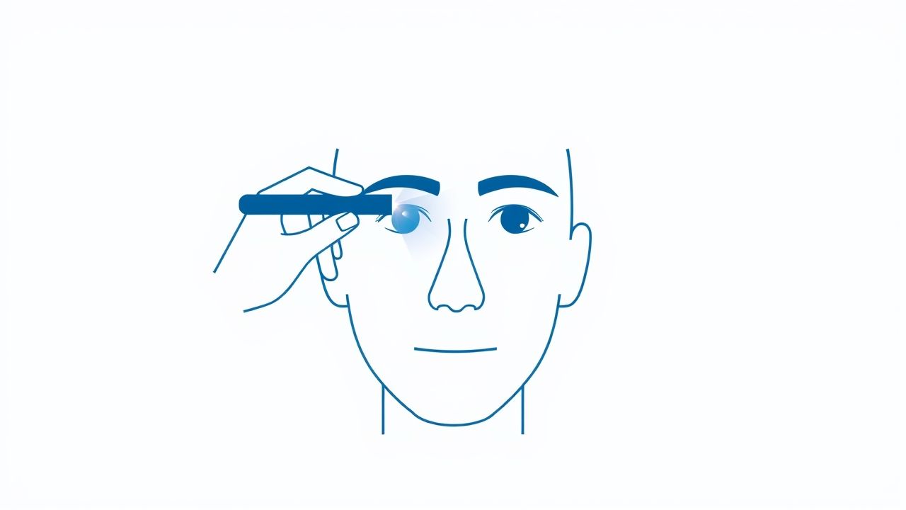
The main goal of using a penlight to check pupils is to observe how they react to changes in light. Pupil size and reaction speed can indicate whether the eyes and brain are functioning normally.
Neurological Health
The pupils are directly connected to the brain through the optic nerve and the oculomotor nerve. Changes in reaction can point to neurological issues that may require further investigation.
Eye Health
Pupil examination can help detect problems such as optic nerve damage, eye trauma, or certain diseases that affect vision.
Medical Emergencies
In emergency settings, checking pupils with a penlight is often one of the first neurological assessments performed to detect possible head injuries, strokes, or drug effects.
How the Penlight Test Works
The penlight pupil check works by stimulating the eye with a direct beam of light, which should trigger a reflex reaction known as the pupillary light reflex.
Pupillary Light Reflex
When light enters the eye, the pupil constricts to limit the amount of light reaching the retina. This reflex is automatic and controlled by the nervous system.
Direct and Consensual Response
- Direct response– The pupil in the eye being tested should constrict immediately when exposed to light.
- Consensual response– The pupil in the opposite eye should also constrict at the same time, even though the light is not shining directly into it.
Step-by-Step Procedure
Although simple, the pupil check should follow a consistent method to ensure accurate results.
Preparation
- Ensure the room has moderate or dim lighting to make pupil changes more visible.
- Ask the person to look at a fixed point in the distance to reduce eye movement.
- Hold the penlight in your dominant hand for better control.
Performing the Test
- Stand slightly to the side of the person being tested to avoid blocking their vision.
- Shine the penlight into one eye for about one to two seconds.
- Observe the size change in the illuminated pupil (direct response).
- Immediately look at the other eye to check for simultaneous constriction (consensual response).
- Repeat the process on the other eye.
Normal Findings
Healthy pupils are usually round, equal in size, and respond quickly to light.
Typical Pupil Size
In normal light conditions, pupil size typically ranges from 2 to 4 millimeters. In darkness, they dilate to 4 to 8 millimeters.
Expected Reaction
Both pupils should constrict rapidly and evenly when exposed to light and return to their previous size when the light is removed.
Abnormal Findings and Their Possible Causes
Variations in pupil size or reaction may indicate underlying health issues.
Unequal Pupil Size (Anisocoria)
- May be harmless in some individuals but can also indicate nerve damage or brain injury.
- Can result from conditions like Horner’s syndrome or third cranial nerve palsy.
Sluggish Reaction
- Can be caused by certain medications, eye diseases, or brain injury.
- May indicate increased intracranial pressure in emergency situations.
No Reaction to Light
- May result from severe optic nerve damage, retinal detachment, or brain death in critical cases.
- Can also occur after certain types of eye surgery.
Fixed and Dilated Pupils
- Often seen in severe head trauma or after cardiac arrest.
- May be linked to drug overdose involving stimulants or hallucinogens.
Factors That Can Affect the Test
Not all unusual pupil reactions indicate a medical emergency. Various factors can temporarily influence results.
Medications
Eye drops for glaucoma, certain painkillers, or recreational drugs can cause dilation or constriction without indicating permanent damage.
Lighting Conditions
Pupils naturally adjust based on environmental lighting, so testing should be done in a controlled setting.
Age
Older adults may have slower pupillary responses due to natural aging of the nervous system.
Clinical Uses Beyond Neurology
Checking pupils with a penlight is not only useful for detecting neurological problems but also in other areas of medicine.
Ophthalmology
Eye doctors use pupil responses to detect optic nerve issues, cataracts, or retinal problems.
Anesthesia Monitoring
During surgery, anesthesiologists observe pupil size as part of monitoring a patient’s depth of anesthesia.
Toxicology
Emergency physicians check pupils when assessing possible drug intoxication or poisoning.
Tips for Accurate Penlight Examination
- Use a focused beam rather than a diffuse light for clearer results.
- Avoid shining the light for too long, as it can cause discomfort and inaccurate readings.
- Compare both eyes carefully to detect subtle differences.
- Document findings immediately if used in a clinical setting.
When to Seek Further Medical Evaluation
If abnormal pupil reactions are observed, it is important to determine whether the cause is urgent.
Emergency Signs
- Sudden onset of unequal pupils after head trauma.
- Pupils that do not respond to light at all.
- Accompanied symptoms like severe headache, confusion, or vision loss.
Non-Emergency Concerns
- Gradual changes in pupil size without other symptoms.
- Persistent anisocoria without an obvious cause.
Checking pupils with a penlight is a simple yet valuable method for assessing eye health and neurological function. By observing how pupils respond to light, medical professionals can detect issues ranging from mild eye conditions to life-threatening brain injuries. While not all abnormal findings indicate an emergency, any unexpected results should be evaluated by a healthcare provider to rule out serious conditions. This quick test remains an essential part of physical examinations and emergency care due to its ability to provide immediate insight into a person’s neurological status.