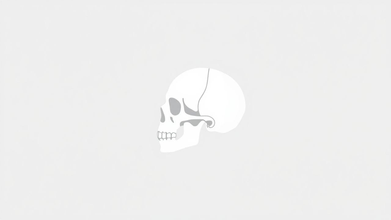
The bones of the fetal skull are fascinating structures that play a crucial role in protecting the developing brain and allowing for safe passage during birth. Unlike the rigid adult skull, the fetal skull is composed of multiple soft bones connected by flexible connective tissue, making it adaptable and capable of growth. This unique arrangement supports brain development, accommodates changes in head size, and facilitates childbirth. Understanding the anatomy, structure, and functions of the bones of the fetal skull provides valuable insights into human development and the remarkable adaptations that occur before birth.
Anatomy of the Fetal Skull
The fetal skull is made up of several individual bones that are not yet fused together. These bones are joined by sutures and fontanelles, which are soft spots allowing flexibility. The major bones include the frontal, parietal, occipital, temporal, sphenoid, and ethmoid bones. These bones form the framework of the head while leaving gaps that enable molding during delivery.
Frontal Bone
The frontal bone forms the forehead and the upper part of the eye sockets. In a fetus, the frontal bone consists of two separate halves divided by a frontal suture. Over time, these halves fuse together, but in early development, the separation helps accommodate rapid brain growth.
Parietal Bones
The two parietal bones are located on each side of the skull, forming most of the top and upper sides. These bones are important in shaping the rounded contour of the fetal head and are connected to other skull bones by sutures, such as the sagittal suture and coronal suture.
Occipital Bone
The occipital bone is positioned at the back and base of the skull. It surrounds the foramen magnum, the large opening through which the spinal cord passes. In fetal development, the occipital bone is divided into several parts that later fuse into one bone during early childhood.
Temporal Bones
The temporal bones are found on the sides of the skull, near the ears. They help form the base of the skull and contain structures essential for hearing and balance. In fetuses, these bones are not completely ossified, which keeps them flexible during birth.
Sphenoid Bone
The sphenoid bone is a central bone at the base of the skull that connects with several other bones. In fetal skulls, parts of the sphenoid remain cartilaginous, allowing for continued growth and adjustment as the brain expands.
Ethmoid Bone
The ethmoid bone is a delicate bone located between the eyes, forming part of the nasal cavity and eye sockets. In fetal skulls, it is small and still developing, but it plays an important role in facial structure.
Sutures of the Fetal Skull
Sutures are fibrous joints that connect the bones of the fetal skull. They allow for movement between bones during birth and provide space for brain growth after birth.
- Coronal suture– between the frontal and parietal bones.
- Sagittal suture– between the two parietal bones.
- Lambdoid suture– between the parietal bones and the occipital bone.
- Metopic suture– between the two halves of the frontal bone.
Fontanelles in the Fetal Skull
Fontanelles, also known as soft spots, are large membrane-covered spaces between the skull bones where sutures meet. They provide extra flexibility for the head to change shape during birth and expand as the brain grows.
- Anterior fontanelle– diamond-shaped and located at the junction of the coronal and sagittal sutures.
- Posterior fontanelle– triangular and located at the junction of the sagittal and lambdoid sutures.
- Sphenoidal fontanelle– located at the junction of the coronal and squamosal sutures.
- Mastoid fontanelle– located at the junction of the lambdoid and squamosal sutures.
Functions of the Bones of the Fetal Skull
The fetal skull’s structure offers multiple advantages that support life before and after birth.
Protection of the Brain
Even though the bones are not fused, the fetal skull still provides a protective casing for the brain. The arrangement of bones and connective tissue cushions the brain against mild pressures.
Facilitation of Childbirth
During delivery, the unfused bones and flexible sutures allow the head to change shape slightly, making it easier for the baby to pass through the birth canal. This process is known as molding.
Support for Brain Growth
Since the brain continues to grow rapidly during infancy, the open sutures and fontanelles give the skull room to expand without causing damage to the brain tissues.
Differences Between Fetal and Adult Skull Bones
The fetal skull differs from the adult skull in several ways. The bones are softer, more numerous, and connected by wider sutures. The presence of fontanelles is a key difference, as adult skulls do not have these soft spots. Additionally, the proportion of the cranium to the face is larger in fetuses, reflecting the early growth of the brain compared to facial bones.
Development and Ossification
Ossification of the fetal skull bones occurs gradually, starting in the second month of fetal life. Most bones form through intramembranous ossification, where bone tissue develops directly from connective tissue membranes. However, parts of the skull base undergo endochondral ossification, where bone replaces cartilage. Fusion of sutures and closure of fontanelles occur over months to years after birth.
Clinical Importance
Examining the bones of the fetal skull can provide important diagnostic clues for pediatricians. Delayed closure of fontanelles, abnormal head shapes, or premature fusion of sutures (craniosynostosis) can indicate developmental concerns or medical conditions that require attention.
Common Conditions Affecting the Fetal Skull
- Craniosynostosis– premature fusion of one or more sutures, leading to abnormal skull shapes.
- Hydrocephalus– excessive accumulation of cerebrospinal fluid causing an enlarged head.
- Microcephaly– abnormally small head size due to limited brain growth.
The bones of the fetal skull are remarkable in their design and function, combining flexibility with protection to support both development and the birthing process. From the frontal bone to the occipital bone, each part plays a vital role in shaping the head and safeguarding the brain. Understanding their anatomy, growth patterns, and clinical significance provides insight into the complexity of human development and the delicate balance between structure and function in early life.