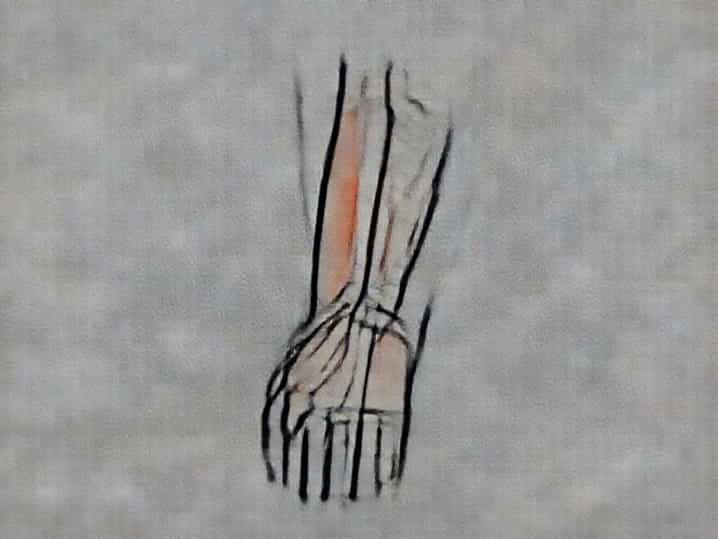
The anterior compartment of the forearm is a crucial anatomical region that plays a significant role in the movement and function of the wrist, hand, and fingers. This compartment houses muscles, nerves, and blood vessels that enable flexion, pronation, and fine motor skills essential for daily activities. Understanding its anatomy, function, and clinical relevance is important not only for medical professionals but also for anyone interested in human physiology, sports science, or rehabilitation. A detailed exploration of the anterior compartment of the forearm highlights its structure, subdivisions, and significance in maintaining upper limb functionality.
Overview of the Anterior Compartment
The anterior compartment of the forearm is located on the front or palmar side of the forearm, extending from the elbow to the wrist. It is primarily responsible for flexion movements of the wrist and fingers, as well as pronation, which rotates the forearm so the palm faces downward. The compartment is encased by fascia, separating it from the posterior compartment, and contains a combination of superficial and deep muscles, along with associated nerves and blood vessels.
Fascial Boundaries
The anterior compartment is enclosed by the antebrachial fascia, which provides structural support and separates it from surrounding tissues. This fascia helps contain the muscles and neurovascular structures, maintaining organized function. Any swelling or trauma within this compartment can increase pressure, potentially leading to conditions such as compartment syndrome, which requires prompt medical attention.
Muscular Composition
The muscles in the anterior compartment of the forearm are primarily flexors and pronators. They are divided into superficial, intermediate, and deep layers, each with distinct roles and attachments. These muscles originate from the medial epicondyle of the humerus or nearby bones and insert into the wrist, hand, or fingers.
Superficial Muscles
The superficial layer consists of muscles that mainly flex the wrist and fingers
- Flexor carpi radialisFlexes and abducts the wrist.
- Flexor carpi ulnarisFlexes and adducts the wrist.
- Palmaris longusFlexes the wrist and tenses the palmar aponeurosis.
- Pronator teresPronates the forearm.
- Flexor digitorum superficialisFlexes the middle phalanges of the fingers.
Intermediate Muscle
The flexor digitorum superficialis sometimes is considered part of an intermediate layer because it lies between the superficial and deep muscles. It plays a vital role in flexing the proximal interphalangeal joints and contributes to grip strength and hand dexterity.
Deep Muscles
The deep layer includes muscles responsible for more refined finger movements and pronation
- Flexor digitorum profundusFlexes the distal phalanges of the fingers.
- Flexor pollicis longusFlexes the thumb, crucial for pinching and grasping.
- Pronator quadratusPronates the forearm, working in conjunction with the pronator teres.
Nerve Supply
The anterior compartment of the forearm receives innervation primarily from the median and ulnar nerves. The median nerve supplies most of the superficial and deep flexors, including the flexor digitorum superficialis, flexor pollicis longus, and pronator muscles. The ulnar nerve specifically innervates the flexor carpi ulnaris and the medial portion of the flexor digitorum profundus. This dual innervation ensures coordinated control of wrist and finger movements, allowing precise and powerful actions.
Blood Supply
The compartment is vascularized mainly by branches of the radial and ulnar arteries, which provide oxygen and nutrients essential for muscle function. The radial artery travels along the lateral aspect, while the ulnar artery supplies the medial structures. Proper circulation is critical for endurance and strength, and any vascular compromise can impact hand and forearm performance.
Functions of the Anterior Compartment
The anterior compartment of the forearm performs multiple essential functions
Wrist Flexion and Stabilization
The flexor carpi radialis, flexor carpi ulnaris, and palmaris longus flex the wrist, contributing to hand positioning during tasks such as typing, gripping, or lifting. They also stabilize the wrist to allow effective force transmission from the forearm to the fingers.
Finger Flexion
Flexor digitorum superficialis and flexor digitorum profundus enable bending of the fingers at the proximal and distal interphalangeal joints, respectively. These movements are crucial for gripping objects, manipulating tools, and performing fine motor tasks like writing or buttoning clothes.
Thumb Flexion and Opposition
Flexor pollicis longus flexes the thumb and assists in opposition movements, enabling precision grip. The thumb’s unique mobility, supported by these muscles, allows humans to perform complex tasks requiring dexterity.
Pronation of the Forearm
Pronator teres and pronator quadratus rotate the forearm so that the palm faces downward. This movement is essential for activities such as turning doorknobs, using utensils, or performing sports actions that require pronation.
Clinical Relevance
The anterior compartment of the forearm is clinically significant due to its involvement in several conditions and injuries. Understanding its anatomy is important for diagnosis, treatment, and rehabilitation.
Compartment Syndrome
Swelling or bleeding within the compartment can increase pressure, leading to compartment syndrome. This condition can compromise blood flow and nerve function, potentially causing muscle necrosis and permanent disability if not treated promptly. Early recognition and surgical intervention through fasciotomy are critical.
Tendinitis and Overuse Injuries
Repetitive use of the flexor muscles, common in typing, sports, or manual labor, can lead to tendinitis. Symptoms include pain, swelling, and reduced grip strength. Proper ergonomics, rest, and physiotherapy are important for recovery.
Nerve Compression
Median or ulnar nerve compression in the forearm can lead to sensory disturbances, weakness, and impaired motor function. Conditions such as pronator syndrome involve compression of the median nerve by the pronator teres, causing pain and numbness in the hand.
Exercises and Strengthening
Strengthening the muscles of the anterior compartment improves grip, wrist stability, and forearm endurance. Exercises include wrist curls, finger flexion with resistance bands, and pronation/supination drills. Regular stretching and strengthening reduce the risk of injury and enhance performance in sports and daily activities.
Practical Tips
- Warm up and stretch the forearm muscles before physical activity
- Use proper ergonomics during repetitive tasks
- Incorporate resistance training to strengthen flexor and pronator muscles
- Monitor for pain or swelling and seek medical advice if symptoms persist
The anterior compartment of the forearm is a vital anatomical region responsible for wrist and finger flexion, forearm pronation, and precise hand movements. Composed of layered muscles, nerves, and blood vessels, it ensures functional dexterity and strength. Awareness of its structure, functions, and potential clinical issues is essential for healthcare providers, athletes, and anyone performing repetitive hand tasks. Understanding this compartment enhances appreciation of the forearm’s complexity and the remarkable coordination required for everyday activities, from gripping objects to performing intricate manual tasks. Proper care, exercise, and awareness of common injuries help maintain its functionality and prevent long-term complications, highlighting the significance of the anterior compartment in human anatomy.