The adrenal medulla is a vital component of the adrenal gland, playing a central role in the body’s response to stress by producing catecholamines such as adrenaline and noradrenaline. Understanding its embryological origin provides key insights into its structure, function, and developmental biology. The adrenal medulla’s origin is distinct from that of the adrenal cortex, which highlights the complexity of adrenal gland development and the specialized cellular lineage that contributes to its unique endocrine functions. Studying the embryological background helps medical professionals and researchers better understand congenital adrenal disorders and the physiological adaptations associated with stress response.
Overview of the Adrenal Gland
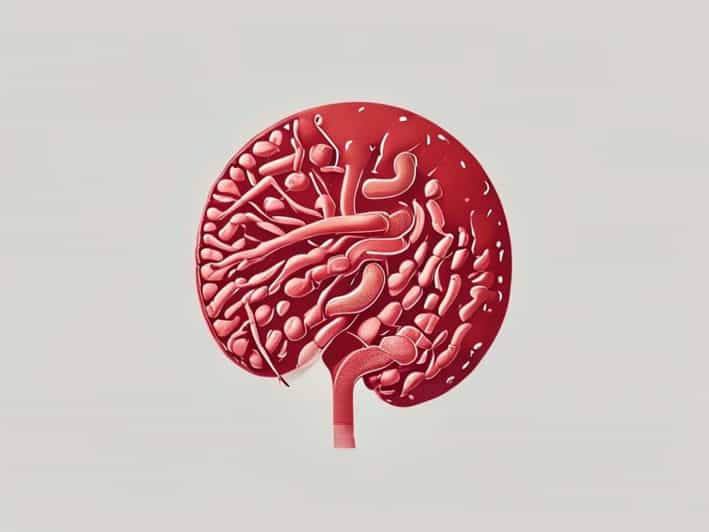
The adrenal glands are paired endocrine organs located atop each kidney. Each gland consists of two main components the outer cortex and the inner medulla. The cortex is responsible for producing steroid hormones such as cortisol, aldosterone, and androgens, whereas the medulla synthesizes catecholamines critical for immediate stress responses. The embryological origins of these two regions differ significantly, reflecting their unique functions and developmental pathways. While the cortex arises from mesodermal tissue, the medulla originates from neural crest cells, emphasizing the dual lineage of the adrenal gland.
Structural and Functional Significance of the Medulla
The adrenal medulla constitutes the inner portion of the adrenal gland and is composed of chromaffin cells, which secrete adrenaline and noradrenaline. These catecholamines are essential for the fight or flight response, increasing heart rate, blood pressure, and blood glucose levels. From an embryological perspective, the medulla’s neural crest origin explains its neuroendocrine properties, as it shares similarities with sympathetic ganglia. Understanding its development is crucial for clinicians dealing with pheochromocytomas, congenital adrenal hyperplasia, and other disorders related to adrenal function.
Embryological Development of the Adrenal Medulla
The adrenal medulla originates from neural crest cells, a group of multipotent cells that emerge from the dorsal neural tube during early embryogenesis. These cells migrate extensively and differentiate into various cell types, including neurons, glial cells, and chromaffin cells of the adrenal medulla. The migration process is tightly regulated by molecular signals and growth factors, ensuring that neural crest cells reach the developing adrenal cortex, where they form the medulla. This developmental pathway underscores the close relationship between the medulla and the sympathetic nervous system.
Neural Crest Cell Migration
During embryogenesis, neural crest cells undergo an epithelial-to-mesenchymal transition, allowing them to migrate to distant locations. For the adrenal medulla, these cells travel along specific pathways from the neural tube to the dorsal mesentery near the developing kidneys. Once they reach the adrenal primordium, interactions with mesodermal cells of the developing cortex guide their differentiation into functional chromaffin cells. Disruptions in neural crest migration can lead to congenital defects affecting the medulla, highlighting the importance of precise embryological development.
Cellular Differentiation of the Medulla
Upon reaching the adrenal cortex, neural crest cells undergo differentiation under the influence of signaling molecules such as glucocorticoids produced by the surrounding cortical cells. These signals promote the maturation of chromaffin cells, which acquire the capacity to synthesize catecholamines. This process illustrates the interdependence between the cortex and medulla during development. By understanding these differentiation pathways, researchers gain insights into how the adrenal medulla achieves its neuroendocrine functions and how developmental disruptions may result in clinical disorders.
Role of Growth Factors and Signaling Molecules
- Glucocorticoids from the adrenal cortex stimulate the conversion of neural crest cells into functional chromaffin cells.
- Bone morphogenetic proteins (BMPs) and fibroblast growth factors (FGFs) regulate proliferation and migration of neural crest cells.
- Transcription factors such as Phox2b and Mash1 guide chromaffin cell specification and maturation.
Timeline of Adrenal Medulla Development
The adrenal medulla begins to develop around the fifth week of gestation, shortly after the formation of the adrenal cortex from mesodermal tissue. By the seventh to eighth week, neural crest-derived cells start migrating into the adrenal primordium. Differentiation into chromaffin cells is largely complete by the tenth week, coinciding with the establishment of functional catecholamine synthesis. This developmental timeline ensures that by birth, the adrenal medulla is capable of responding to stress signals, although full maturation and regulation continue postnatally.
Functional Maturation
Although structural differentiation occurs prenatally, the functional maturation of the adrenal medulla continues after birth. Catecholamine secretion is present at birth, but regulatory mechanisms, such as response to sympathetic stimulation, develop gradually. For medical professionals, understanding both the structural and functional aspects of medullary development is crucial for diagnosing and managing neonatal adrenal disorders and stress-related conditions.
Clinical Implications of Embryological Origin
The neural crest origin of the adrenal medulla has significant clinical implications. Disorders arising from defective neural crest migration, such as congenital central hypoventilation syndrome or neuroblastoma, often involve the adrenal medulla. Neuroblastoma, a cancer of neural crest-derived cells, frequently affects the adrenal medulla in children, underscoring the importance of embryological knowledge for early detection and treatment. Additionally, understanding the medulla’s development helps explain certain congenital adrenal hyperplasias and their impact on catecholamine production and stress response.
Disorders Linked to Developmental Abnormalities
- Pheochromocytoma Tumor of chromaffin cells affecting catecholamine secretion.
- Neuroblastoma Malignant tumor originating from undifferentiated neural crest cells in the adrenal medulla.
- Congenital adrenal hyperplasia May indirectly affect medullary function due to cortical-medullary interactions.
- Neural crest migration defects Lead to structural and functional abnormalities of the adrenal medulla.
Comparative Embryology
Studying the adrenal medulla across species provides additional insights into its evolution and functional specialization. In vertebrates, the adrenal medulla consistently originates from neural crest cells, while variations exist in its size, chromaffin cell distribution, and hormonal output. Comparative studies highlight the conserved mechanisms of neural crest migration, differentiation, and adrenal integration, providing valuable information for developmental biology and translational research.
The adrenal medulla’s embryological origin from neural crest cells is a critical aspect of its development, structure, and function. Its migration, differentiation, and interaction with the adrenal cortex underscore the complex processes that create a functional neuroendocrine organ. Understanding the developmental timeline, signaling pathways, and clinical implications of medullary formation allows for better diagnosis, treatment, and management of adrenal disorders. For medical professionals, researchers, and students of developmental biology, the study of the adrenal medulla provides a compelling example of how specialized cell lineages contribute to essential physiological functions. From catecholamine synthesis to stress response, the embryological foundation of the adrenal medulla remains central to its role in human health and disease.