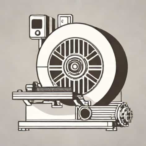
X-ray imaging has been a cornerstone of medical diagnostics and material analysis for over a century, allowing non-invasive visualization of internal structures. While traditional X-ray techniques rely primarily on absorption contrast, advancements in imaging technology have led to the development of methods that enhance sensitivity to subtle structural differences. One such method is X-ray imaging using a grating interferometer, which allows for phase-contrast imaging and differential phase measurements. This technique significantly improves the visualization of soft tissues and fine structures that are difficult to observe with conventional absorption-based X-rays, expanding applications in medicine, biology, and materials science. Understanding how grating interferometers work, their components, and their advantages provides insight into why this method is becoming increasingly important in modern imaging.
Principles of Grating Interferometer X-ray Imaging
A grating interferometer utilizes the wave nature of X-rays to extract additional information beyond conventional absorption. Unlike standard X-ray imaging that detects the intensity reduction as X-rays pass through a material, grating interferometry measures the phase shift induced by the sample. This phase shift corresponds to the refractive index variations within the object, revealing structural details with higher contrast and sensitivity.
Basic Components of a Grating Interferometer
Grating interferometers typically consist of three main components
- Source Grating (G0)Used in setups with conventional X-ray tubes to create an array of individually coherent X-ray beamlets.
- Phase Grating (G1)Introduces a periodic phase modulation to the incoming X-ray beam, creating an interference pattern downstream.
- Analyzer Grating (G2)Positioned at the detector side to convert phase variations into detectable intensity changes, allowing quantitative measurement of phase shifts.
These gratings are carefully aligned, and the setup may include additional components like precision stages for scanning and positioning. The precise spacing and orientation of the gratings determine the sensitivity and resolution of the imaging system.
How Grating Interferometry Works
When X-rays pass through a sample, their phase is altered depending on the refractive index and thickness of the material. The phase grating causes these shifts to produce an interference pattern, which is then analyzed by the analyzer grating. By performing a process known as phase-stepping, the system captures multiple images with small lateral shifts of the grating, allowing extraction of three types of contrast information
- Absorption contrastConventional attenuation of X-ray intensity.
- Differential phase contrastSensitivity to small angular deviations of X-rays caused by the sample.
- Dark-field contrastInformation about microstructural variations and scattering properties.
These three complementary contrasts enable detailed visualization of both dense and soft tissues, as well as internal microstructures of materials, providing a multidimensional perspective that is not possible with standard X-ray imaging.
Applications in Medical Imaging
Grating interferometer X-ray imaging has significant potential in clinical and preclinical settings. One of the main advantages is its ability to improve the visualization of soft tissues without the need for contrast agents. This can be especially beneficial for early detection of diseases such as tumors, pulmonary disorders, and cartilage degeneration.
Soft Tissue Imaging
Traditional X-ray imaging struggles to distinguish between soft tissues like muscles, fat, and internal organs due to similar absorption properties. Phase-contrast imaging enhances contrast between these tissues by measuring refractive index differences, providing clearer images for diagnosis and research.
Lung and Breast Imaging
Research has shown that grating interferometry can detect subtle structures in lung tissue, which can improve early diagnosis of conditions like emphysema or fibrosis. Similarly, in mammography, phase-contrast X-rays can enhance the visibility of microcalcifications and tumors, potentially improving cancer detection rates while reducing the radiation dose compared to traditional mammography.
Applications in Materials Science
Beyond medicine, grating interferometer X-ray imaging is widely used in materials science for non-destructive testing and quality control. The ability to detect fine structural variations and microcracks in metals, ceramics, and composites is valuable in research, aerospace, and engineering applications.
Microstructure Analysis
By examining differential phase and dark-field contrasts, scientists can study the internal fiber orientation, porosity, and microstructural defects of materials. This technique allows for a detailed understanding of material behavior under stress and during manufacturing processes, leading to better quality control and performance assessment.
Non-destructive Testing
Grating interferometer X-ray imaging enables inspection of components without causing damage. For example, in aerospace engineering, it can be used to detect internal flaws in turbine blades, composite panels, and other critical parts. The combination of absorption, phase, and dark-field information provides a comprehensive assessment of structural integrity.
Advantages of Grating Interferometer Imaging
The use of a grating interferometer in X-ray imaging offers several notable advantages
- Enhanced soft tissue contrast compared to conventional X-rays.
- Ability to detect microstructural and sub-resolution features using dark-field imaging.
- Quantitative phase information that can be analyzed for material properties or tissue density.
- Compatibility with standard X-ray sources, enabling easier integration into existing imaging setups.
- Potential for dose reduction while maintaining image quality, particularly in medical applications.
Challenges and Limitations
Despite its advantages, grating interferometer X-ray imaging also has challenges that researchers and practitioners must consider. The precision required in grating fabrication and alignment can be technically demanding. High-quality gratings are expensive and sensitive to environmental conditions such as vibration and temperature fluctuations.
Field of View and Scan Time
Another limitation is the restricted field of view, which can require multiple scans for large samples or patients. Phase-stepping procedures can also increase scan time compared to conventional X-ray imaging, although advancements in fast detectors and automated systems are mitigating this issue.
Accessibility
Currently, widespread clinical adoption is limited by the complexity and cost of the technology. Research institutions and specialized imaging centers are the primary users, though ongoing developments aim to create more compact and cost-effective systems suitable for routine clinical use.
Future Directions
Grating interferometer X-ray imaging continues to evolve, with ongoing research focusing on improving sensitivity, reducing scan time, and increasing accessibility. Hybrid imaging systems that combine conventional absorption with phase and dark-field information are being developed to provide comprehensive diagnostic data. Additionally, integration with computed tomography (CT) techniques enables three-dimensional visualization of phase and dark-field contrasts, further expanding applications in medicine and materials science.
Integration with AI and Computational Methods
Artificial intelligence and advanced image reconstruction algorithms are being applied to grating interferometry data to enhance contrast, reduce noise, and extract quantitative metrics. These computational techniques will play a critical role in translating this powerful imaging method into practical, real-world applications.
X-ray imaging using a grating interferometer represents a significant advancement in non-invasive imaging technology. By exploiting the phase information and scattering properties of X-rays, this technique provides enhanced soft tissue contrast, microstructural visualization, and multi-contrast imaging that surpass conventional absorption-based methods. Applications in medicine, materials science, and non-destructive testing demonstrate the versatility and value of this approach. Although challenges such as grating precision, field of view, and accessibility remain, ongoing research and technological improvements are paving the way for broader adoption. As phase-contrast and dark-field imaging become more integrated into standard X-ray systems, grating interferometer imaging promises to expand the capabilities of diagnostic imaging and material analysis, providing more detailed and informative views of the internal structure of objects and tissues.