When people look for pictures of muscle atrophy in legs, they are often trying to understand how this condition appears in real life and what visual signs indicate weakening muscles. Muscle atrophy can develop gradually or suddenly depending on the cause, and noticing early changes can be important for recognizing underlying issues. While no images are shown here, exploring common visual characteristics, causes, and progression helps create a clear mental picture of what leg muscle atrophy looks like and how it affects the body. Understanding these details can also help people identify when to seek medical guidance or begin appropriate rehabilitation.
What Muscle Atrophy in the Legs Looks Like
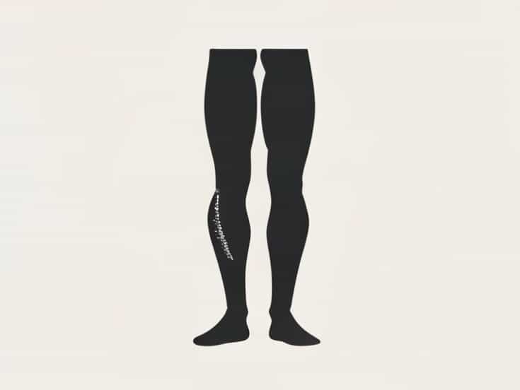
Muscle atrophy refers to the shrinking or wasting away of muscle tissue. In the legs, it often becomes noticeable because the limbs lose fullness and definition. When describing pictures of muscle atrophy in legs, the most common features include decreased muscle mass, visible contours of bone, and uneven size between legs.
Visible Loss of Muscle Mass
The most defining characteristic is thinning of the muscles. Areas such as the thighs, calves, or even the gluteal region may appear significantly smaller than usual. Someone familiar with their own leg shape might notice that the leg looks flatter or less rounded, especially when compared to the opposite leg if only one side is affected.
Increased Prominence of Bones and Tendons
As muscle tissue decreases, bones and connective structures become more visible. Kneecaps may appear sharper, the shin bone more pronounced, and tendons around the ankle may stand out in ways they did not before. This contrast is often emphasized in medical photos capturing advanced muscle wasting.
Asymmetry Between Legs
One common detail seen in pictures of muscle atrophy in legs is unevenness. A person might have one leg significantly smaller than the other, especially if the cause is related to injury, nerve damage, or immobilization of one limb. This asymmetry is often a key indicator of localized atrophy.
Causes of Muscle Atrophy in the Legs
Understanding the causes helps people interpret what they see in photos and descriptions. Several conditions or situations can lead to muscle loss in the legs, each producing its own pattern of visual change.
Disuse or Immobilization
One of the most common causes is lack of movement. When a leg is in a cast, or when someone has been bedridden for long periods, muscles weaken rapidly. Pictures of muscle atrophy in legs due to disuse often show dramatic thinning of the calf and thigh after weeks or months without weight-bearing activity.
Nerve Damage
Nerve injuries or neurological disorders can also lead to atrophy because muscles rely on nerve signals to function. Conditions such as peripheral neuropathy, spinal cord injury, or nerve compression may produce noticeable shrinking. In such cases, atrophy can occur even if the person tries to use the affected muscles.
Chronic Illness or Systemic Conditions
Some diseases cause overall muscle wasting. Conditions such as muscular dystrophy, ALS, or severe malnutrition may lead to progressive thinning across both legs. Photos showing this type of atrophy often reveal symmetrical loss of mass and reduced strength.
Aging
Age-related muscle loss, known as sarcopenia, develops gradually. Visual signs may include softer or looser skin around the legs and less muscle definition. Though not as dramatic as other causes, pictures of older adults with sarcopenia show clear long-term thinning of leg muscles.
Stages Often Seen in Leg Muscle Atrophy
Descriptions of images commonly highlight the stages of atrophy, helping people understand how the condition progresses.
Early Stage
- Slight decrease in muscle definition
- Reduced firmness of the thighs or calves
- Mild decrease in strength
In early pictures of muscle atrophy in legs, changes can be subtle and easily overlooked.
Moderate Stage
- Visible reduction in size of affected muscles
- Increased visibility of bone structure
- Noticeable asymmetry if one leg is more affected
At this stage, differences become much easier to identify, even without close examination.
Advanced Stage
- Significant wasting of muscle groups
- Very prominent bones and tendons
- Difficulty standing, walking, or bearing weight
Photos of advanced leg muscle atrophy often show stark thinning and require medical attention to identify the underlying condition.
How People Interpret Photos of Leg Muscle Atrophy
Medical images can be challenging to interpret for someone without experience. Understanding common visual patterns can make descriptions clearer.
Comparing Before-and-After Views
Side-by-side comparisons are often used to demonstrate progression or improvement. These pictures highlight how quickly muscle can deteriorate when untreated and how rehabilitation restores volume.
Focusing on Specific Muscle Groups
Leg atrophy can affect
- Quadriceps (front of the thigh)
- Hamstrings (back of the thigh)
- Calves
- Hip stabilizers
Pictures that emphasize specific muscle groups help identify which areas have weakened and may guide physical therapy approaches.
Distinguishing Atrophy From Weight Loss
General weight loss affects fat and muscle, while atrophy specifically affects muscle tissue. Descriptions of pictures of muscle atrophy in legs emphasize tighter skin against deeper structures, while general weight loss is more uniform.
What Pictures Cannot Show
While images provide visual cues, they cannot show pain levels, cause, or functional limitations. Someone with mild-looking atrophy may struggle significantly with mobility, while someone with dramatic visible muscle loss may function better than expected due to compensatory strength elsewhere.
Symptoms Not Visible in Photos
- Weakness when climbing stairs
- Fatigue in the legs after short activity
- Numbness or tingling from nerve issues
- Instability or difficulty balancing
These symptoms help complete the picture beyond what photographs alone can capture.
Reversibility of Leg Muscle Atrophy
Many people look for pictures of muscle atrophy in legs to understand whether recovery is possible. In many cases, regaining muscle mass is achievable with the right approach.
Exercise and Physical Therapy
Targeted strengthening exercises can rebuild lost muscle. Visual progress often shows fuller muscle shape and improved symmetry over time. Therapists frequently document changes to track rehabilitation.
Nutrition Support
Protein intake, essential nutrients, and proper hydration support muscle repair. While not visible directly in photos, these factors influence the pace of visual improvement.
Treating Underlying Conditions
If nerve damage or illness causes atrophy, treating the root issue is key. Pictures showing recovery frequently follow successful treatment plans that address more than just the muscle itself.
Although no pictures are displayed here, understanding the typical visual features of muscle atrophy in legs helps create a clear mental image of how the condition appears. Thinning muscle mass, noticeable bone structure, and asymmetry are common indicators seen in medical descriptions and photographs. Recognizing these signs is important for identifying potential causes, understanding progression, and knowing when to seek help. With the right combination of treatment, exercise, and support, many people can restore strength and improve the appearance of affected leg muscles over time.