The pleura is a vital anatomical structure within the human respiratory system, yet many people may not be fully aware of what it is or why it matters. Found in the thoracic cavity, the pleura surrounds the lungs and plays a crucial role in breathing, protection, and maintaining a smooth interface between organs. Understanding the pleura helps explain not only how the lungs work efficiently but also what happens when diseases affect this area, such as pleurisy or pleural effusion. Whether you’re a student of medicine, a healthcare professional, or simply curious about the body, a clear explanation of the pleura is essential for understanding respiratory anatomy.
Definition and Function of the Pleura
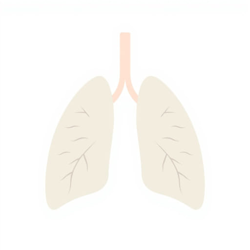
What Is the Pleura?
The pleura is a thin, double-layered membrane that lines the thoracic cavity and covers the lungs. This structure is composed of two layers:
- Visceral pleura: The inner layer that tightly adheres to the surface of the lungs, dipping into the fissures between the lobes.
- Parietal pleura: The outer layer that lines the inner wall of the chest cavity, including the diaphragm and the mediastinum.
Between these two layers is the pleural cavity, a narrow space filled with pleural fluid. This fluid acts as a lubricant, allowing the lungs to expand and contract smoothly during breathing without friction.
Main Functions of the Pleura
The pleura has several essential roles in maintaining respiratory health:
- Reducing friction: The pleural fluid prevents the lungs from rubbing against the chest wall during inhalation and exhalation.
- Creating surface tension: This helps keep the lungs inflated by adhering the lungs to the chest wall, even during changes in pressure.
- Protection: The pleural layers provide a barrier against infections and injury.
Anatomical Structure of the Pleura
Visceral Pleura
The visceral pleura is in direct contact with the lungs and cannot be separated from the lung surface. It follows the contours of the lung lobes and provides a smooth covering that enables effortless movement during respiration.
Parietal Pleura
The parietal pleura is more extensive and attaches to multiple structures in the thoracic cavity, including:
- The inner surface of the rib cage (costal pleura)
- The diaphragm (diaphragmatic pleura)
- The side of the mediastinum (mediastinal pleura)
- The apex of the lung area near the neck (cervical pleura)
Each of these sections has unique relationships with nearby organs and nerves, which is important in clinical diagnosis and surgery.
Pleural Cavity and Pleural Fluid
The Pleural Space
The pleural cavity is not a true open space but rather a potential space between the visceral and parietal pleura. Under normal conditions, this cavity contains a small amount of serous fluid typically around 10 to 20 milliliters.
Pleural Fluid
The pleural fluid serves a critical purpose in reducing friction and helping the lungs maintain their position against the chest wall. It also facilitates efficient gas exchange by ensuring that the lungs remain expanded with minimal energy use.
Too much or too little pleural fluid, however, can lead to complications such as pleural effusion (fluid buildup) or pneumothorax (air in the pleural space).
Clinical Significance of the Pleura
Pleural Diseases and Disorders
Because of its location and function, the pleura can be involved in several medical conditions. These include:
- Pleuritis or pleurisy: Inflammation of the pleura, often causing sharp chest pain that worsens during breathing.
- Pleural effusion: Accumulation of excess fluid in the pleural space, which can be caused by infection, heart failure, or cancer.
- Pneumothorax: Air enters the pleural cavity, causing the lung to collapse. This can result from trauma or spontaneously in certain individuals.
- Hemothorax: Accumulation of blood in the pleural space, often due to injury or surgery.
- Mesothelioma: A rare and aggressive cancer affecting the pleura, commonly linked to asbestos exposure.
Diagnosis and Treatment
Diagnosing pleural conditions typically involves a combination of imaging techniques and clinical tests:
- Chest X-raysandCT scansare used to visualize fluid, air, or abnormalities.
- Pleural fluid analysisthrough thoracentesis helps identify infections, cancer, or other causes of effusion.
- Ultrasoundcan guide fluid drainage and evaluate pleural thickening or adhesions.
Treatment depends on the underlying cause but may involve medications, drainage procedures, or even surgery in severe cases.
Embryological Development of the Pleura
The pleura develops during the early stages of fetal growth. The lungs begin forming as outgrowths of the foregut and push into the surrounding coelomic cavity. As this occurs, the developing lungs become enveloped by a layer of mesoderm that differentiates into the visceral and parietal pleura.
Understanding this development explains why the pleura is intimately associated with both the lungs and the thoracic walls. Any defects during this process can lead to congenital abnormalities affecting breathing and lung function.
Pleura in Other Species
While this topic focuses on human anatomy, it’s worth noting that most mammals have pleural membranes similar in structure and function. Veterinary medicine also studies the pleura to diagnose and treat respiratory issues in animals such as dogs, cats, and horses. The same principles apply: the pleura supports smooth lung motion, prevents damage from friction, and can be affected by trauma or disease.
Protecting Pleural Health
Lifestyle Considerations
Protecting your lungs and pleura begins with overall respiratory health. Here are some simple tips to maintain a healthy pleural environment:
- Avoid smoking: Smoking damages lung tissue and can lead to chronic inflammation or cancer involving the pleura.
- Limit exposure to toxins: Substances like asbestos, silica, and industrial chemicals can harm the pleura.
- Stay physically active: Regular exercise improves lung function and circulation, benefiting pleural health.
- Get vaccinations: Infections such as pneumonia or tuberculosis can spread to the pleura if untreated.
The pleura may be a thin membrane, but its role in respiratory anatomy is both complex and essential. Comprising two layers the visceral and parietal pleura it ensures that the lungs function efficiently by minimizing friction and helping maintain lung inflation. The pleural cavity and its fluid allow the lungs to glide smoothly within the chest during each breath. From inflammation and infections to serious conditions like pneumothorax and mesothelioma, the pleura is clinically significant. Understanding what the pleura is, how it works, and how to care for it can offer valuable insights into both normal anatomy and disease. Whether for education, diagnosis, or general interest, the study of the pleura deepens our appreciation for the intricacies of the human body.