The cardiograph is a fundamental medical device used to monitor and record the electrical activity of the heart. Understanding its function and significance is crucial for both healthcare professionals and individuals interested in cardiovascular health. By providing detailed insights into heart function, the cardiograph helps detect abnormalities, diagnose cardiac conditions, and guide treatment plans. With advancements in technology, modern cardiographs have become more precise, user-friendly, and accessible, making them an essential tool in both hospitals and outpatient settings.
What a Cardiograph Measures
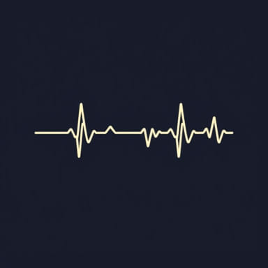
A cardiograph primarily measures the electrical impulses generated by the heart as it contracts and relaxes. These impulses reflect the heart’s rhythm, rate, and electrical conduction patterns. The device translates these signals into a visual graph known as an electrocardiogram (ECG or EKG), which healthcare providers interpret to assess cardiac health. The cardiograph does not directly measure mechanical functions like blood flow or heart muscle contraction but provides indirect insight into these functions through electrical activity patterns.
Heart Rate
One of the most basic measurements obtained from a cardiograph is heart rate. The heart rate represents the number of heartbeats per minute and serves as a primary indicator of cardiac function. A normal adult resting heart rate typically ranges between 60 and 100 beats per minute. Deviations from this range, such as tachycardia (rapid heart rate) or bradycardia (slow heart rate), can signal underlying cardiac conditions that require further investigation.
Heart Rhythm
The cardiograph also measures the rhythm of the heart, indicating whether the heartbeat is regular or irregular. A consistent rhythm, known as sinus rhythm, reflects healthy heart function, whereas irregular rhythms may indicate arrhythmias. Common arrhythmias detected using a cardiograph include atrial fibrillation, atrial flutter, and ventricular tachycardia. Identifying abnormal rhythms is vital because some arrhythmias can lead to serious complications such as stroke or heart failure if left untreated.
Electrical Conduction Patterns
Another critical measurement provided by a cardiograph is the electrical conduction pattern within the heart. The cardiograph captures the timing and sequence of electrical impulses as they travel through different regions of the heart, including the atria and ventricles. Abnormal conduction patterns may indicate issues such as heart block, bundle branch block, or pre-excitation syndromes. By analyzing these patterns, healthcare providers can pinpoint specific areas of the heart that may require intervention.
Components of a Cardiograph
Modern cardiographs consist of several essential components that work together to capture accurate heart measurements. Understanding these components helps in appreciating how the device functions and the quality of data it produces.
Electrodes
Electrodes are small, adhesive sensors placed on the patient’s skin to detect electrical signals from the heart. The placement of electrodes follows specific configurations, such as the standard 12-lead setup, to capture comprehensive data from multiple angles. Proper electrode placement is crucial for obtaining accurate readings and avoiding signal interference.
Amplifier
The electrical signals generated by the heart are extremely weak. The amplifier component of a cardiograph strengthens these signals, making them readable and suitable for recording. Amplification ensures that even minor electrical variations are captured, which is essential for diagnosing subtle cardiac abnormalities.
Recorder or Display
The recorder translates the amplified electrical signals into a visual format, usually a graph called an electrocardiogram. Traditional cardiographs print the ECG on paper, while modern digital cardiographs display it on screens for real-time monitoring. This visual representation allows healthcare providers to interpret the heart’s rhythm, rate, and conduction patterns quickly and accurately.
Types of Cardiographs
Cardiographs have evolved over time, offering different types suited for various clinical scenarios. Each type has unique advantages and is selected based on the patient’s needs and monitoring duration.
Resting Cardiograph
Resting cardiographs are used when the patient is at rest, typically lying down. This type is ideal for routine checkups or initial evaluations of heart function. It provides a snapshot of the heart’s electrical activity under baseline conditions, helping to detect anomalies that may not be apparent during physical activity.
Holter Monitor
The Holter monitor is a portable cardiograph that records the heart’s electrical activity continuously over 24 to 48 hours or longer. It is particularly useful for detecting intermittent arrhythmias or symptoms that occur sporadically. Patients carry the device while engaging in normal daily activities, allowing healthcare providers to assess how the heart functions under various conditions.
Stress Test Cardiograph
A stress test cardiograph monitors the heart’s electrical activity while the patient engages in physical exercise, usually on a treadmill or stationary bike. This test evaluates how the heart responds to increased workload and can reveal conditions such as ischemia or exercise-induced arrhythmias. It provides critical information that resting measurements alone may not detect.
Clinical Importance of Cardiograph Measurements
The measurements obtained from a cardiograph are indispensable in diagnosing, managing, and preventing cardiovascular diseases. They offer insights that guide treatment decisions and improve patient outcomes.
Early Detection of Cardiac Conditions
Cardiographs enable early detection of heart conditions, including arrhythmias, heart block, and myocardial infarction. Early identification allows timely intervention, which can prevent complications and improve long-term health.
Monitoring Treatment Effectiveness
Patients undergoing treatment for heart conditions, such as medication or pacemaker therapy, can benefit from cardiograph monitoring. Regular ECGs help assess the effectiveness of treatment, enabling adjustments to optimize cardiac health and reduce risks.
Pre-Surgical Assessment
Before surgical procedures, a cardiograph is often used to evaluate a patient’s heart function. Detecting underlying cardiac issues ensures that appropriate precautions are taken during surgery, minimizing the risk of complications.
The cardiograph measures the electrical activity of the heart, providing critical information on heart rate, rhythm, and conduction patterns. By capturing these measurements, it serves as a cornerstone in diagnosing, monitoring, and managing cardiovascular diseases. From resting cardiographs to Holter monitors and stress test devices, these tools help healthcare providers make informed decisions and improve patient outcomes. Understanding the role and function of the cardiograph emphasizes its value in modern medicine and highlights the importance of regular cardiac monitoring for maintaining heart health. With technological advancements, cardiographs continue to evolve, offering more precise, accessible, and user-friendly options for patients and healthcare professionals alike.